Figure 3. Slit-lamp photomicrograph of two individuals with Fleck corneal dystrophy. A: In proband 1, punctate discrete opacities are visible in retroillumination (arrows), and a few larger opacities are seen
in the posterior stroma in direct illumination (arrowheads). B: In proband 2, discrete punctate grayish-white opacities are seen diffusely distributed in the corneal stroma (arrows).
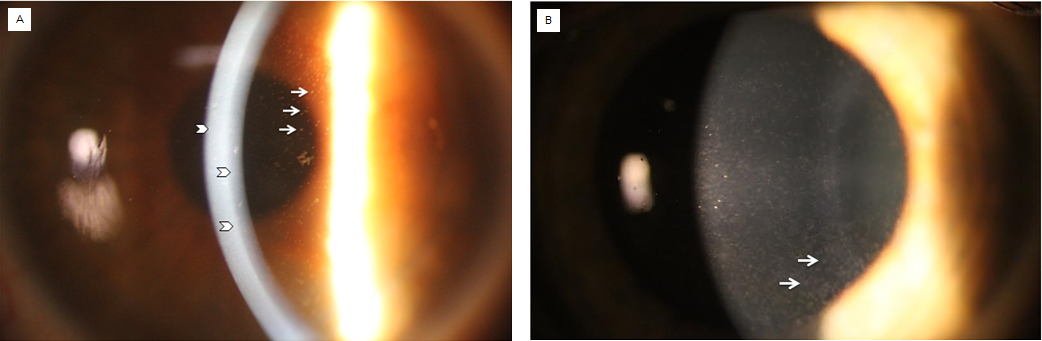
 Figure 3 of
Gee, Mol Vis 2015; 21:1093-1100.
Figure 3 of
Gee, Mol Vis 2015; 21:1093-1100.