Figure 4. Immunohistochemistry for VNUT in C57BL/6J and DBA/2J retinas. Vertical sections at 3, 9, 15, and 22 months of retinas from
glaucomatous mice (B, C, E, F) and control mice at 3 and 15 months (A, D). Vesicular nucleotide transporter (VNUT)-stained retinas (green) showed a significant increase at 15 months in the DBA/2J
mice (E) compared to that observed in the DBA/2J and C57BL/6J mice at 3 months (A, B) and 23 months (F). G–H: High magnification of the photoreceptor layer, showing an increase in VNUT labeling in the DBA/2J mice at 15 months (G) and VNUT staining found in ganglion cells (arrows; H). Nuclei were stained with propidium iodide (red). All images were collected from the central area of the retina. ONL, outer
nuclear layer; OPL, outer plexiform layer; INL, inner nuclear layer; IPL, inner plexiform layer; GCL, ganglion cell layer.
Scale bar: 20 μm.
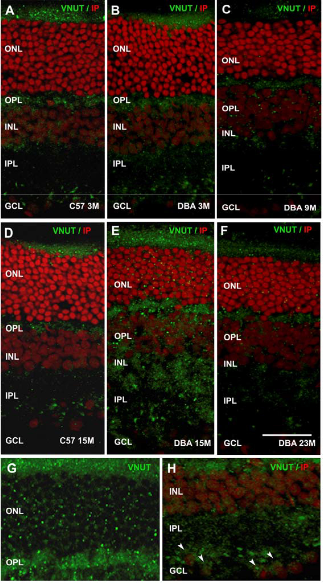
 Figure 4 of
Pérez de Lara, Mol Vis 2015; 21:1060-1070.
Figure 4 of
Pérez de Lara, Mol Vis 2015; 21:1060-1070.