Figure 2. Western blot analysis of β-catenin in HLE-B3 cells treated with SB216763. HLE-B3 cells were cultured in 25 cm2 flasks with 20% fetal bovine serum (FBS) and switched to serum-free media for 24 h before the experiment. The cells were
incubated with 3 ml of serum-free media containing 12 µm SB216763 or SB216763 combined with either 0.5 µm hypoxia-inducible
factor-1α (HIF-1α) translation inhibitor (KC7F2) or HIF-2α translation inhibitor (CAS882268–69–1) for 3 h in hypoxia (1% oxygen).
Cytoplasmic and nuclear lysates were collected from HLE-B3 cell cultures after treatments using the NE-PER Nuclear and Cytoplasmic
Extraction Kit. A portion of the sample was used for protein quantification using the EZQ Protein Quantification Kit, and
3X sodium dodecyl sulfate (SDS) buffer was added to the remaining lysates, which were subsequently boiled for 5 min; the proteins
were resolved by electrophoresis on 12% SDS-polyacrylamide gels (20 μg protein/lane). The proteins were then transferred to
nitrocellulose membranes. The experiment was repeated twice with independent cell populations and the image density of β-catenin
was quantified using ImageJ analysis. The β-catenin levels in the cytoplasmic extracts were essentially unchanged, while in
the nuclear extracts there was a significant increase in β-catenin in the SB216763-treated cells, as well as SB treatment
with the HIF translation inhibitors, compared with the controls. SB=SB216763.
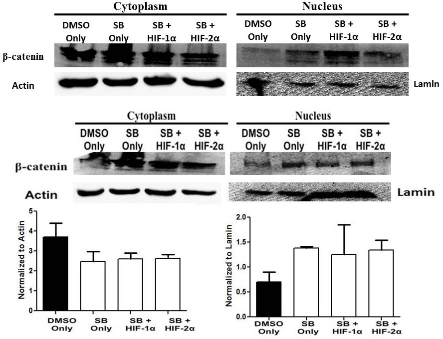
 Figure 2 of
Cammarata, Mol Vis 2015; 21:1024-1035.
Figure 2 of
Cammarata, Mol Vis 2015; 21:1024-1035.