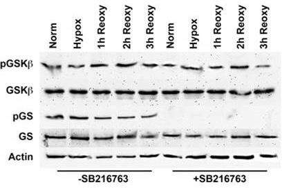
Figure 1. Western blot analysis of GSK-3β and GS phosphorylation in HLE-B3 cells in the presence or absence of SB216763. Total cell
lysates were collected from >85% confluent HLE-B3 cell cultures that were incubated for 90 min in serum-free minimal essential
media (MEM) containing either 12 µM SB216763 or 0.05% DMSO vehicle. Cells were then exposed to hypoxia for 3 h. At the end
of the incubation period, the hypoxic medium was removed, and fresh, oxygenated serum-free MEM with SB216763 or dimethyl sulfoxide
(DMSO) was added to the cultures. Cells were then placed in atmospheric oxygen for up to 3 h. Cultures were collected after
continuous normoxic exposure (approximately 21% oxygen), hypoxic exposure (approximately 1% oxygen), or after reintroduction
of atmospheric oxygen (approximately 21%) for 1, 2, or 3 h. Total cell lysates were analyzed by immunoblots using 25 µg of
protein per lane. Antiactin was used to normalize the bands to ensure equivalent lane loading. Note: These data were taken
from a prior publication [
9] but are typical of SB216763 treatment. Note to reader: Look at the hypoxic exposure lanes to verify that glycogen synthase,
a substrate of glycogen synthase kinase-3β (GSK-3β) treated with SB216763 fails to be phosphorylated. This is indicative of
inactivation of GSK-3β catalytic activity that also prevents phosphorylation of β-catenin.
 Figure 1 of
Cammarata, Mol Vis 2015; 21:1024-1035.
Figure 1 of
Cammarata, Mol Vis 2015; 21:1024-1035.