Figure 1. Ophthalmologic features of the Vogt-Konayagi-Harada (VKH) patient. A, B: Fundus shows bilateral exsudative retinal detachments. C, D: The fluorescein angiogram reveals bilateral multifocal areas of pinpoint leakage at the level of retinal pigment epithelium
and optic nerve staining. E, F: Fundus photograph obtained 1 year later shows a moderate sunset-glow fundus with diffuse depigmentation of retinal pigment
epithelium in both eyes.
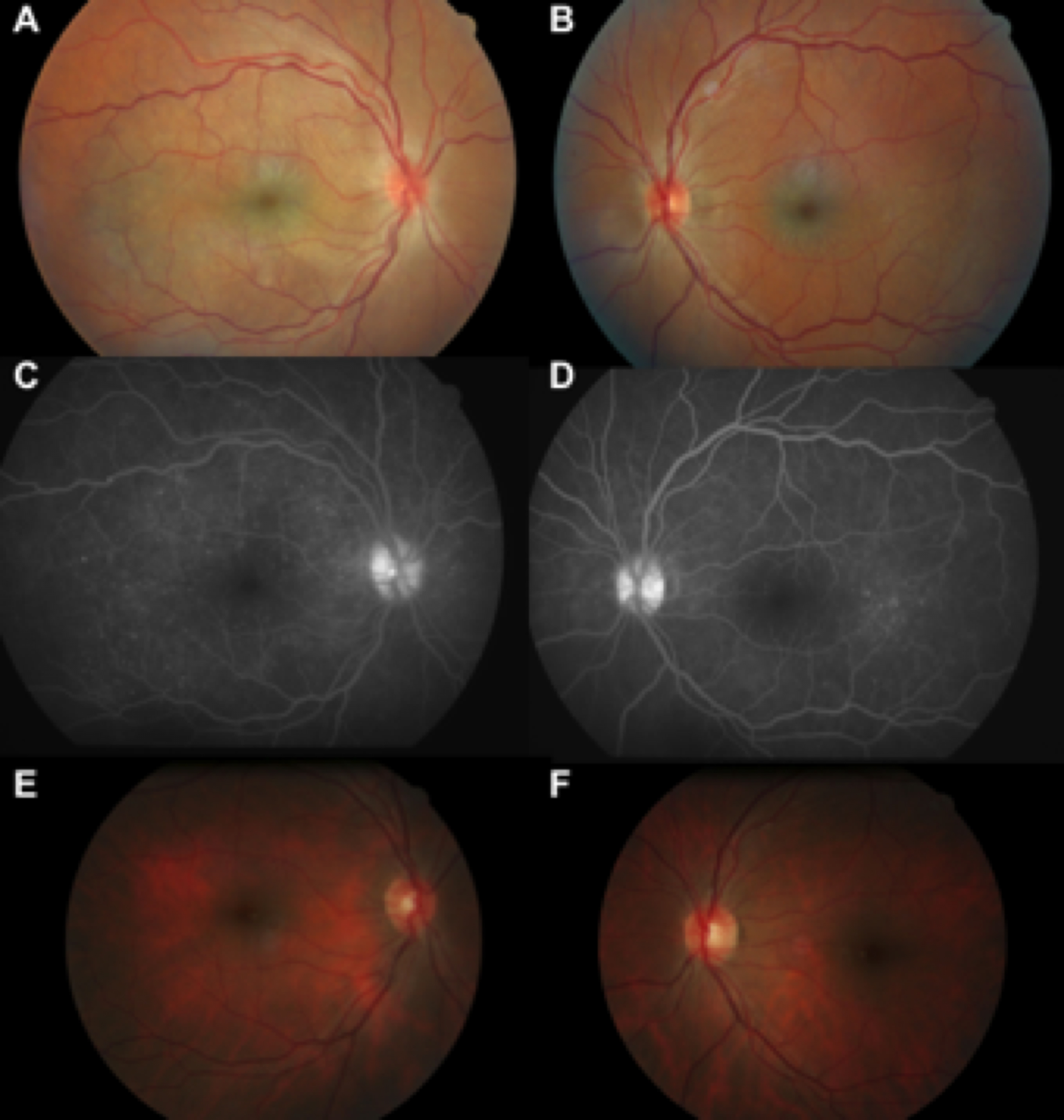
 Figure 1 of
Abad, Mol Vis 2014; 20:956-969.
Figure 1 of
Abad, Mol Vis 2014; 20:956-969.