Figure 2. Anterior segment photographs showing changes in vital staining with 3% topical diquafosol application in the Sod1-/- mice. Upper inserts: Note the improvement in fluorescein staining after diquafosol sodium application. Lower inserts: Note
the improvement in Rose Bengal staining after diquafosol sodium application.
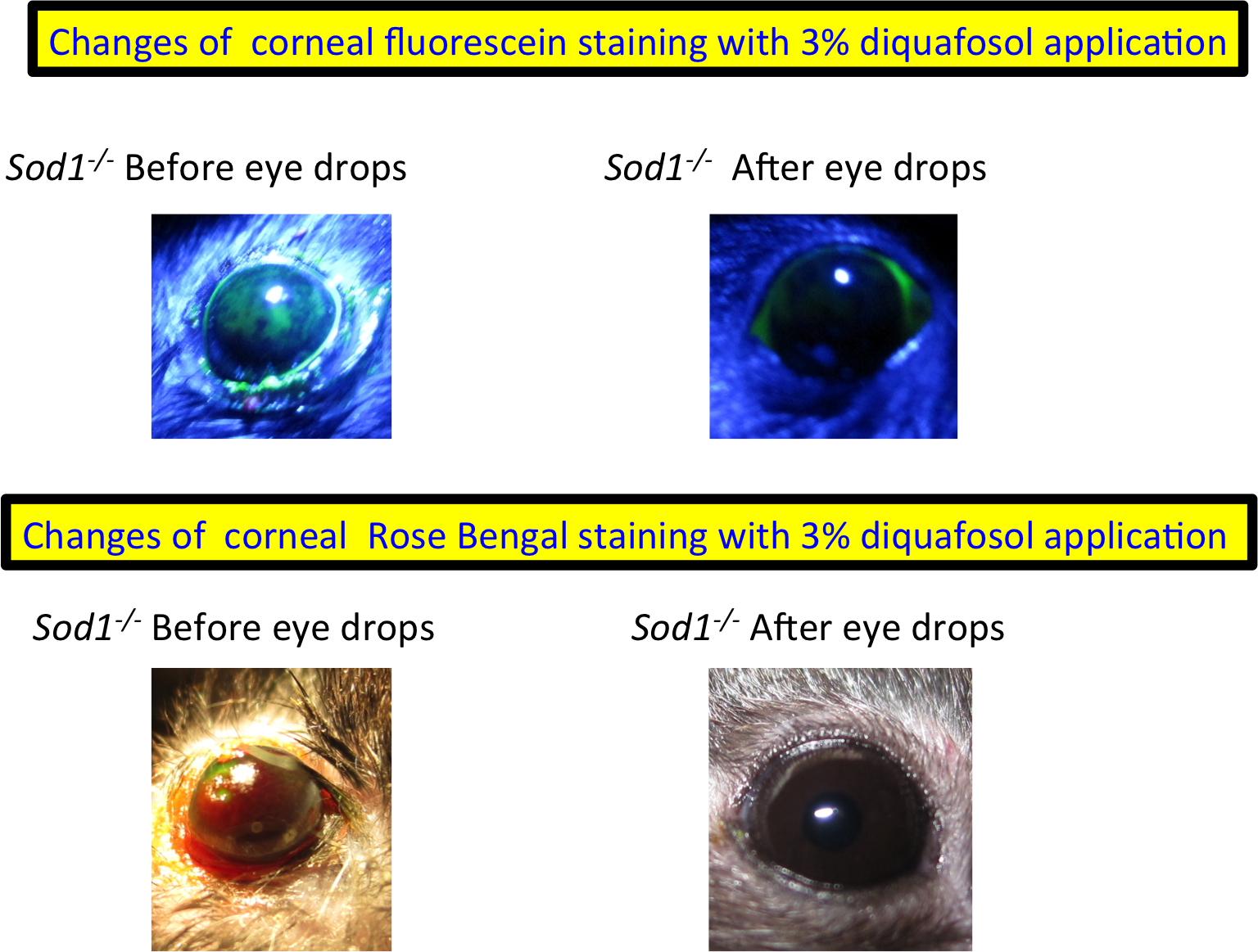
 Figure 2 of
Kojima, Mol Vis 2014; 20:929-938.
Figure 2 of
Kojima, Mol Vis 2014; 20:929-938.