Figure 4. Inducible nitric oxide synthase expression: immunohistochemistry and RT–PCR inducible nitric oxide synthase and ionized calcium-binding
adaptor molecule 1 coimmunostaining on retinal sections of rats with endotoxin-induced uveitis injected with vehicle (A–C) or with antivascular endothelial growth factor (D–F). A and D: Ionized calcium-binding adaptor molecule 1 (IBA1) cells are labeled in green, iNOS is labeled in red and nuclei are stained
in blue with 4’,6-diamidino-2-phenyl-indole (DAPI). B and E: IBA1 cells labeled in green. C and F: Inducible nitric oxide synthase (iNOS) labeled in red. GCL = ganglion cell layer, INL = inner nuclear layer, ONL = outer
nuclear layer, RPE = retinal pigment epithelial cells, Chor: choroid. Bar = 50 µm. Insets are increased magnifications (5X)
of regions delimited by squares. G: iNOS mRNA expression on retinal extracts from vehicle and antivascular endothelial growth factor (anti-VEGF) rat eyes; n
= 5 per groups * p<0.05).
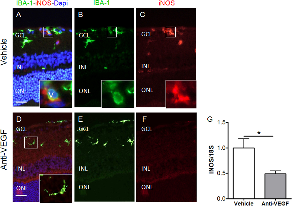
 Figure 4 of
Couturier, Mol Vis 2014; 20:908-920.
Figure 4 of
Couturier, Mol Vis 2014; 20:908-920.