Figure 3. Elevated glucose decreases the interaction between GluA2 subunit and GRIP1 in retinal neural cells. Cells were exposed to
30 mM glucose (Gluc) or 25 mM mannitol (Man - osmotic control; + 5 mM glucose) for 48 h. The interaction between the GluA2
subunit and its interacting proteins was analyzed with coimmunoprecipitation (IP) followed by western blotting (WB). A: Immunoreactivity of GRIP1 and NSF in the immunoprecipitated samples of GluA2. B: Immunoreactivity of GluA2 in the immunoprecipitated samples of GRIP1. C: Immunoreactivity of GluA2 in the immunoprecipitated samples of NSF. The results are presented as the mean ± standard error
of the mean (SEM) of at least three independent experiments and are expressed as a percentage of the control. Representative
immunoblots are presented at the right of the respective graphs. *p<0.05, significantly different from the control as determined
with one-way ANOVA followed by Dunnett’s post hoc test.
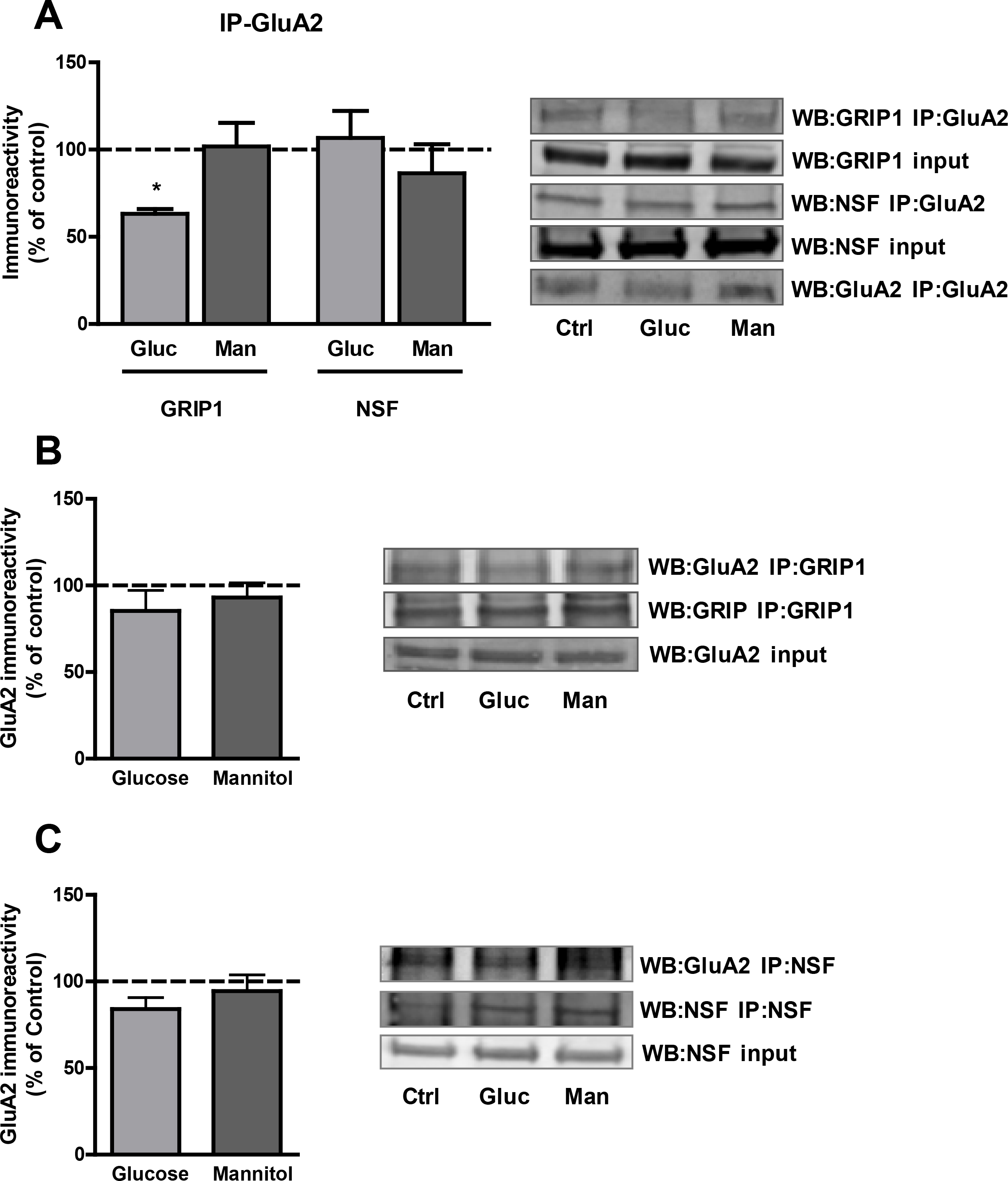
 Figure 3 of
Castilho, Mol Vis 2014; 20:894-907.
Figure 3 of
Castilho, Mol Vis 2014; 20:894-907.