Figure 2. Results of examinations for patients 1Ia and 1IIa. A: The latest Goldmann perimetry shows only small crescent-shaped temporal remnants. B: The fundus photograph shows changes typical for autosomal recessive retinitis pigmentosa (arRP), including pale optic nerve
head, narrowed retinal vessels, bone spicule-like pigmentations in the periphery but also atrophy and pigmentations in the
macula. C–D: Optical coherence tomography (OCT) measurement of macular thickness demonstrates attenuation in all segments of the retinal
thickness map. E: Multifocal electroretinography (mERG) shows severely reduced amplitudes and delayed implicit times compared to a normal
registration (F). G: The fundus photograph from patient 1IIa reveals pigment changes in the macula and midperiphery, deep orange-yellow retinal
flecks in the posterior pole and midperiphery, and a slightly pale optic nerve head. H–I: OCT measurement of retinal thickness shows an attenuated retina in all segments. J: mERG demonstrates severely reduced central and paracentral amplitudes and delayed implicit times.
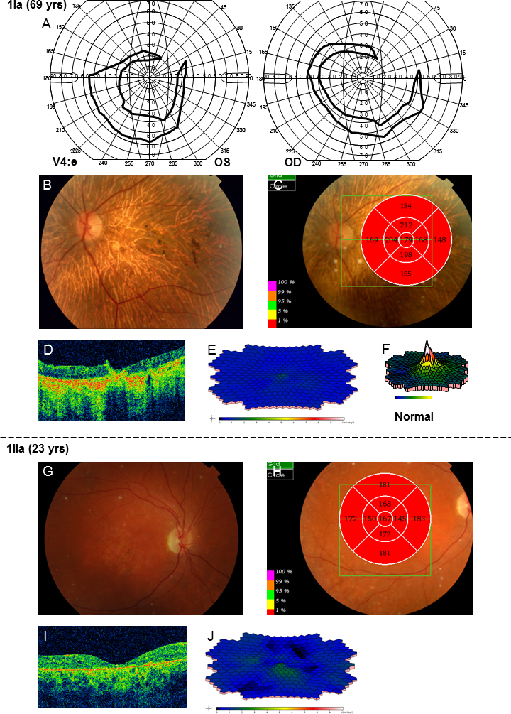
 Figure 2 of
Kjellström, Mol Vis 2014; 20:89-104.
Figure 2 of
Kjellström, Mol Vis 2014; 20:89-104.