Figure 4. Immunohistochemical localization for macrophages in retrolental membranes from eyes with retinopathy of prematurity. A: Expression of CD40 (representing M1 polarity; arrows) and CD206 markers (representing M2 polarity; arrowheads) in tissue
macrophages associated with retinopathy of prematurity (ROP) tissue. B–C: Antihuman inducible nitric oxide synthase (iNOS) and arginase antibodies were also used to detect M1 and M2 macrophages,
respectively. D: Distribution of M1 and M2 macrophages in retrolental membranes from eyes with retinopathy of prematurity (ROP); the predominant
macrophage is the M1 type, and there is only a limited number of M2 type macrophages (** p<0.001). Scale bar = 20 µm.
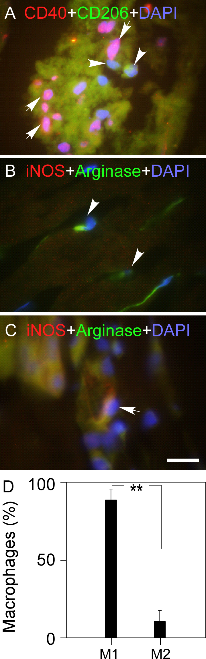
 Figure 4 of
Ma, Mol Vis 2014; 20:881-893.
Figure 4 of
Ma, Mol Vis 2014; 20:881-893.