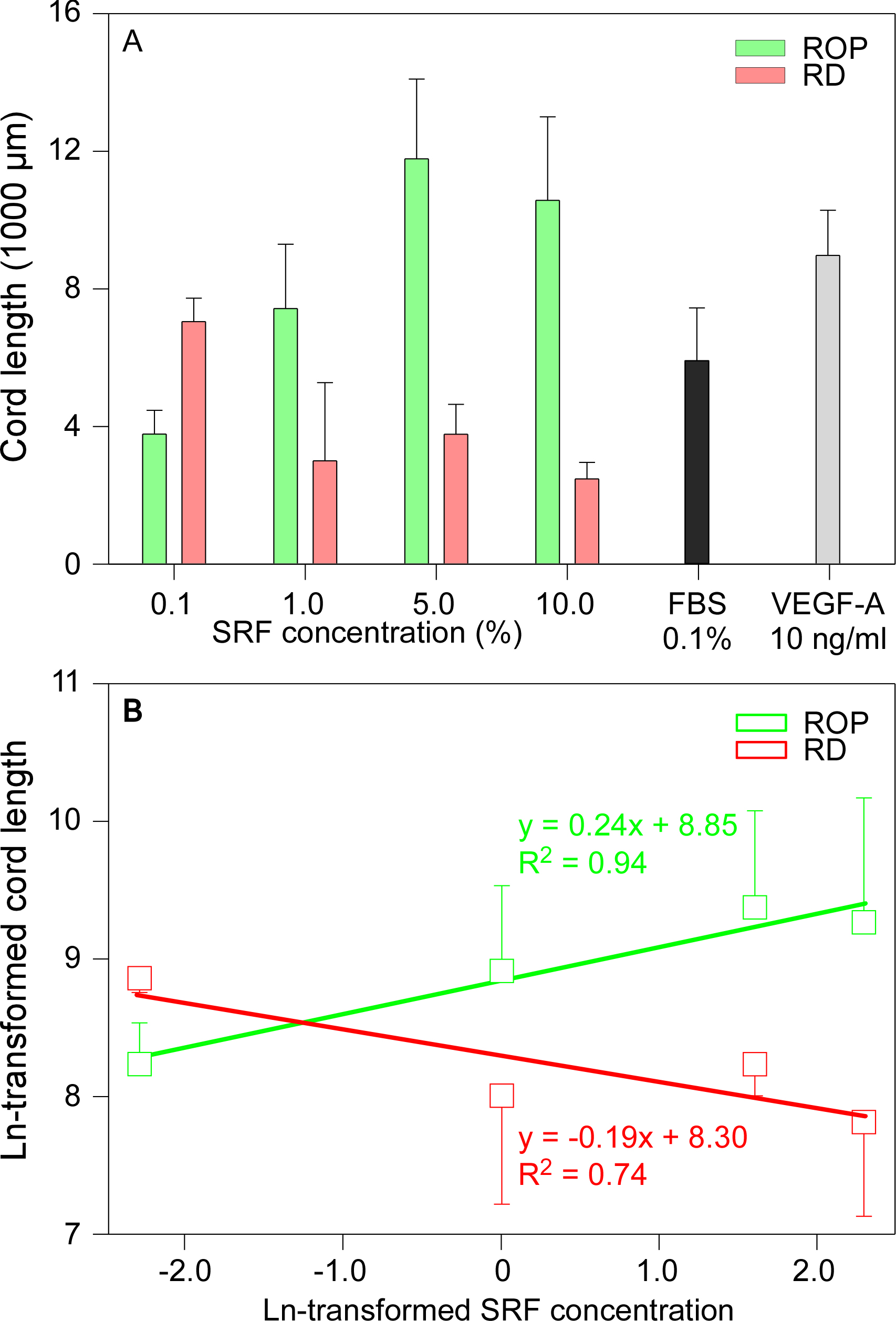
Figure 2. Three-dimensional fibrin clot model of cord formation by human microdermal capillary endothelial cells incubated with subretinal
fluid from eyes with retinopathy of prematurity and retinal detachment. The capillary cord was formed in all culture conditions
and indicates that the cord length from the medium alone was longer than that from cells cultured in a medium containing SRF
from patients with retinal detachment (RD).
A: Subretinal fluid (SRF) from eyes with retinopathy of prematurity (ROP) induced cord formation in human microdermal capillary
endothelial cells (HMCECs) while SRF from eyes with RD showed shorter length measurements (Student t test, n = 10/group, *
p < 0.05, Mean±SEM, please see
Table 2 for detailed comparisons). Vascular endothelial growth factor A (VEGF-A; 10 ng/ml) and 0.1% fetal bovine serum (FBS) were
used as positive and negative controls, respectively.
B: A linear association was found between the cord length and the SRF concentration (ROP, positive; RD, negative). The cord
length and the SRF concentration were natural logarithm (Ln)-transformed to establish the linear models.
 Figure 2 of
Ma, Mol Vis 2014; 20:881-893.
Figure 2 of
Ma, Mol Vis 2014; 20:881-893.