Figure 6. Spectral-domain optical coherence tomogram and fundus autofluorescence in a female carrier of RPGR ORF15 X-linked retinitis pigmentosa. Horizontal transfoveal spectral-domain optical coherence tomogram (A) from the left eye of the female carrier of XLRP after 2 h of dark adaptation at the age of 22 years. An ordered configuration
of the photoreceptor/retinal pigment epithelium complex is seen. Except a slightly undulating ellipsoid line, there seems
to be no obvious difference compared to the normal optical coherence tomogram of a healthy subject. Retina layers from bottom
upward: RPE=retinal pigment epithelium/Bruch’s membrane complex, ISe=inner segment ellipsoid, ONL=outer nuclear layer, OPL=outer
plexiform layer, INL=inner nuclear layer, IPL=inner plexiform layer, NFL=nerve fiber layer. Autofluorescence fundus photography
was performed at the age of 22 years. No abnormal pattern and thus no reminiscence of tapetal-like reflex was noted on imaging
obtained in the light adapted state (B, last image in a series of five images), or after 2 h of dark adaptation (C, first image in a series of five images). The patient’s autofluorescence fundus images displayed a sensitivity to dark adaptation
that is also known from subjects with normal eyes. Thus, a progression was seen from the first to the last image in that the
intensity of autofluorescence gradually increased, except in the fovea. This phenomenon is believed to reflect the bleaching
of the photoreceptor photopigment as the result of continued exposure to blue light and the shielding of the foveal fluorophores
by the xanthophyll pigment. The autofluorescence images showed no trace of the tapetal reflex pattern, neither before nor
after bleaching.
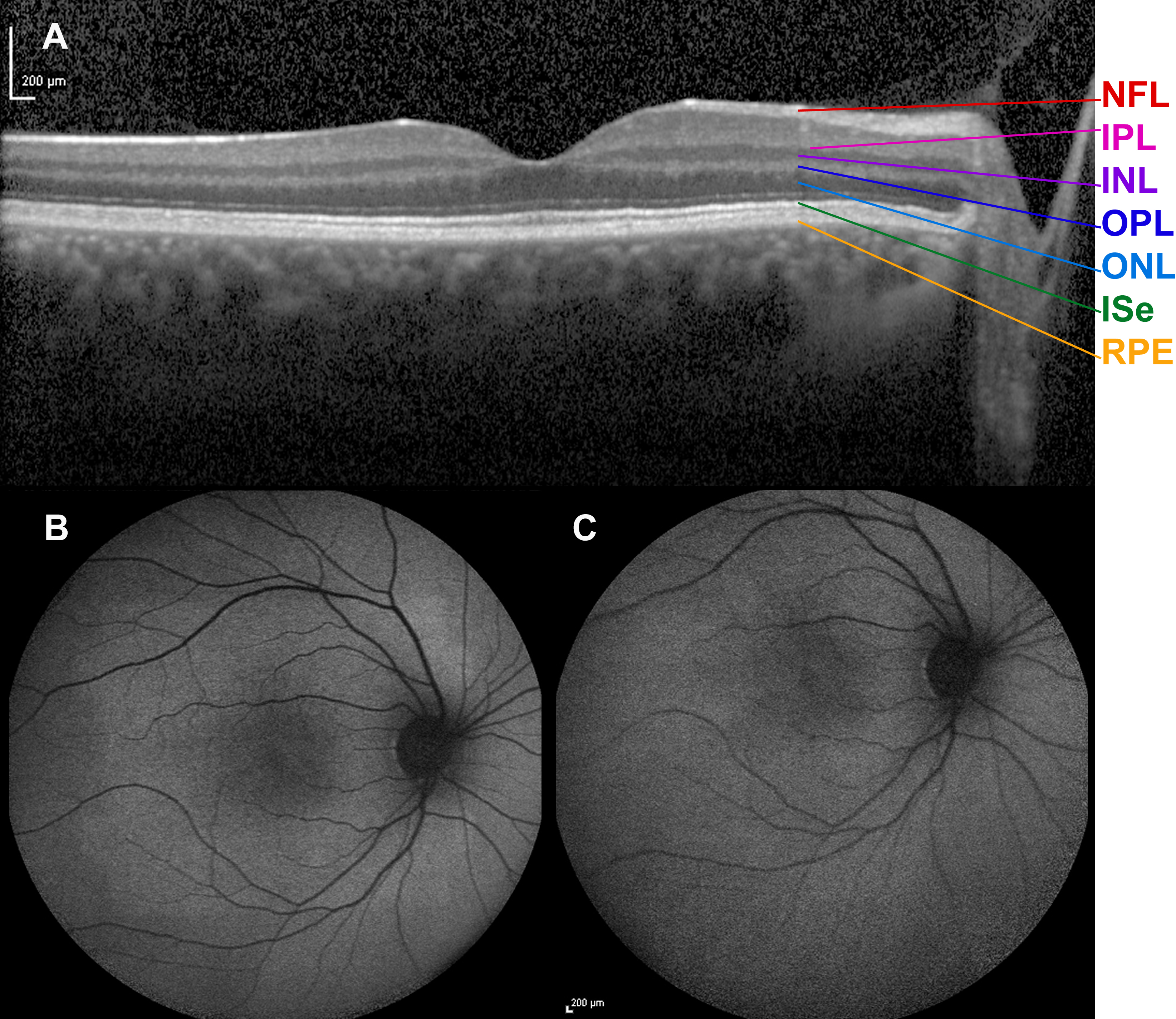
 Figure 6 of
Bregnhøj, Mol Vis 2014; 20:852-863.
Figure 6 of
Bregnhøj, Mol Vis 2014; 20:852-863.