Figure 1. Cosegregation analysis of the identified alleles and schematic representation of EPHA2 wild-type and mutant proteins. A: Pedigree with genotype data for EPHA2, CRYBB3, and CRYBA2 alleles. Individuals affected with congenital cataract are indicated by shaded symbols. Genotyping results for the three
alleles identified in the family are shown below each individual tested: 1 = EPHA2; 2 = CRYBB3; 3 = CRYBA2. The pathogenic allele is indicated in bold. The proband is indicated with an arrow; wt, wild-type allele at the variant position.
B: Schematic drawing of the EPHA2 protein and C-terminal extension mutant sequences. The EPHA2 domain structure is shown at
the top; SP = signal peptide, FN III = fibronectin III type repeats, TM = transmembrane domain, JMR = juxtamembrane region,
SAM = sterile alpha motif, PDZ = PDZ-binding motif. C-terminal sequences of EPHA2 wild-type and frameshift mutants are shown
at the bottom with the PDZ-motif residues indicated in blue and erroneous amino acids in red.
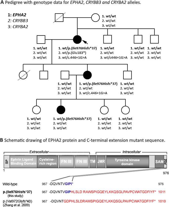
 Figure 1 of
Reis, Mol Vis 2014; 20:836-842.
Figure 1 of
Reis, Mol Vis 2014; 20:836-842.