Figure 7. The atomic structure of the Ca-binding site and the functional consequences of the G1310S mutant variant are shown. Ca-binding
site structures for the wild-type and the mutant variant are shown in beige and light blue, respectively. The Ca2+ ion is shown in green. The oxygen and hydrogen atoms are represented by red and white spheres, respectively.
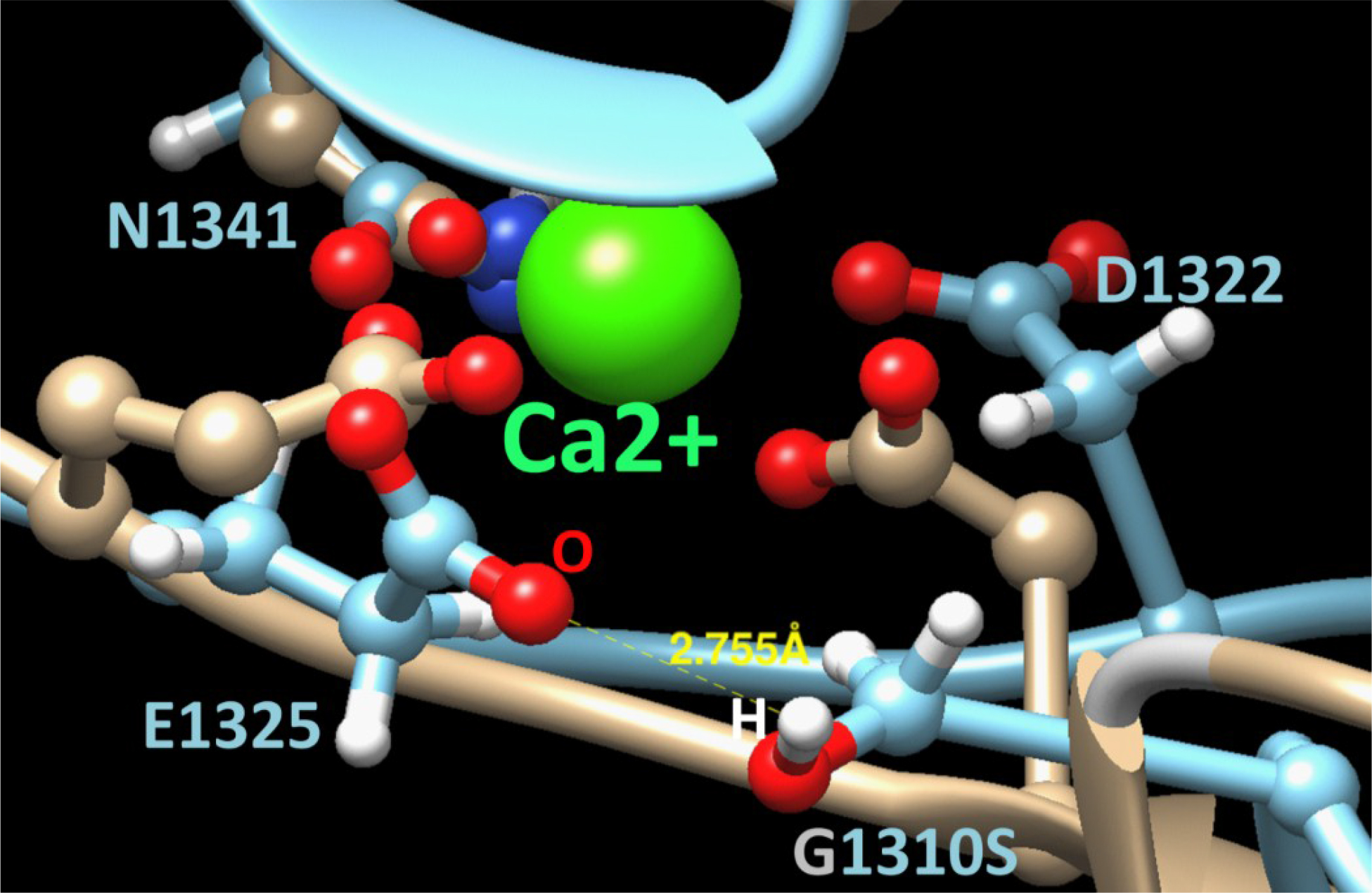
 Figure 7 of
Fu, Mol Vis 2014; 20:812-821.
Figure 7 of
Fu, Mol Vis 2014; 20:812-821.