Figure 3. Laser capture microdissection of limbal crypt cells. A: Diagram depicting the tissue section sandwiched between the polyethylene naphtalate (PEN) membrane and a microscope glass
slide. B: Before the capture, the basal limbal crypt cells (blue) and the corneal cells (green) are identified microscopically based
on their histological structure. C: The basal crypt cells are fixed to the cap by applying infrared (IR) laser pulses (red), melting the cap focally to the
PEN membrane, after which the cells are dissected from the remaining tissue by cutting around the area with an ultraviolet
(UV) laser (blue bars). D: The cap with the cells is lifted from the remaining PEN membrane, the capture of the basal crypts cells is confirmed with
microscopic inspection, and the cells are processed for RNA.
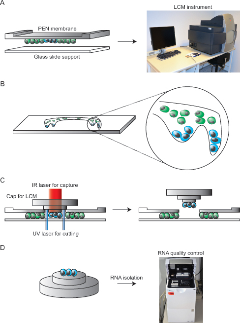
 Figure 3 of
Bath, Mol Vis 2014; 20:797-803.
Figure 3 of
Bath, Mol Vis 2014; 20:797-803.