Figure 1. Preparation of histological sections from cornea. A: The dotted lines in the left panel represent the line of dissection of the bulbus, and the dissected eye segment is shown
on the right. B: The corneal slicer was constructed to feature a Teflon base with a spherical depression, into which the cornea was placed
face down, and the metal frame into which three parallel blades were fixed 4 mm apart. The resulting corneal blocks are shown
on the right. C: Schematic outline of the procedure, including a detail of the corneal limbus from slice stained with cresyl violet. The
asterisk indicates a limbal crypt.
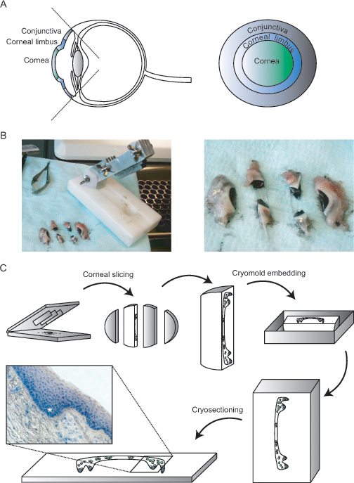
 Figure 1 of
Bath, Mol Vis 2014; 20:797-803.
Figure 1 of
Bath, Mol Vis 2014; 20:797-803.