Figure 3. Clinical evaluations of patients from the eight families with mutations in retinitis pigmentosa genes. A: Patient 01-V:1 (age 22, carrying PRPF31 p.Leu366fs*1) has a waxy optic disc, attenuated retinal arterioles, and pigment deposits around the superior periphery of
the left eye. B: Fundus examination of Patient 02-II:2 (age 22, carrying SNRNP200 p.R681H) shows numerous bone spicule-like pigments and crystalline forms were scattered in the peripheral fundus (arrow).
C: The fundus of Patient 02-I:2 (age 51, carrying SNRNP200 p.R681H) shows intensive pigment deposits. D: Patient 03-IV:3 (age 22, carrying SNRNP200 p.S1087L) demonstrates diffuse bone spicule-like pigment deposits in the periphery retina of the right eye. E: Patient 04-III:3 (age 65, carrying IMPDH1 p.D311N) has intensive bone spicule-like pigment deposits, which severely impacted the central retina. F: The fundus of Patient 05-II:1 (age 58, carrying RP1 p. S781*) shows typical retinitis pigmentosa (RP) features with posterior subcapsular cataract. G: The fundus of Patient 06-II:1 (age 26, carrying RHO p.P347L) with typical RP features. H: Fundus examination of Patient 07-II:1 (age 23, carrying RP2 p.Ile137del) reveals typical features of RP fundus. I: Patient 08-II:5 (age 49, carrying RP2 p.Q65*) has typical fundus of RP. J: Multifocal electroretinogram (mfERG) test traces from the right eye of Patient 02-II:2 are shown. Responses of the peripheral
retina are greatly reduced while responses within the central 10 degrees are slightly involved. K: MfERG of the right eye of Patient 02-I:2 demonstrates a global decrease in ERG responses. L: ERG responses of one unrelated control. M: Optical coherence tomography (OCT) examination of Patient 02-II:2 shows retinal atrophy. N: OCT examination of Patient 02-I:2 shows macular edema and retinal atrophy.
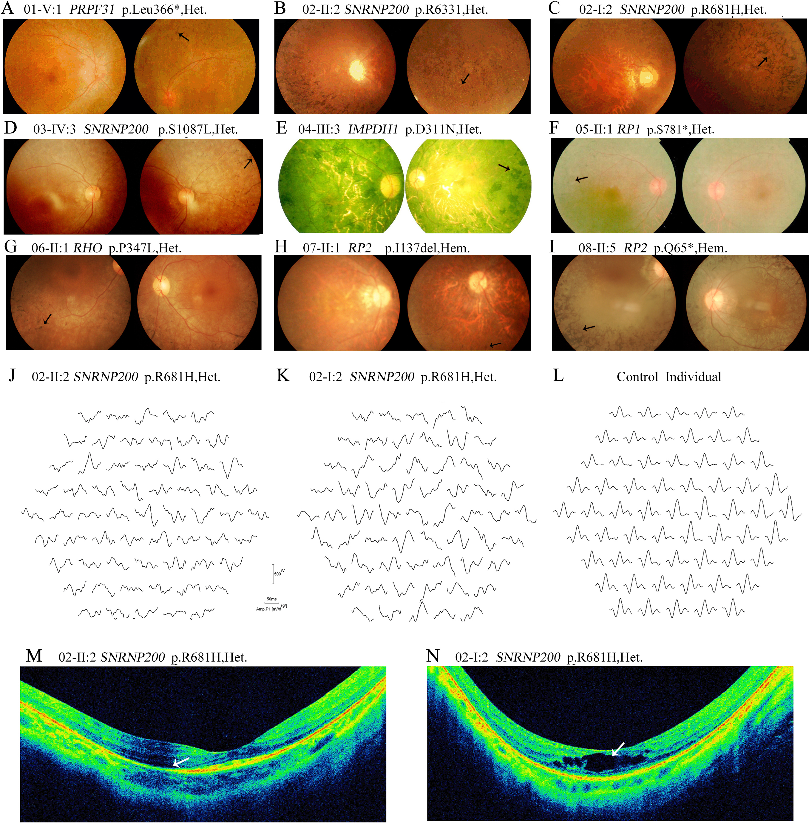
 Figure 3 of
Pan, Mol Vis 2014; 20:770-779.
Figure 3 of
Pan, Mol Vis 2014; 20:770-779.