Figure 5. Retina explants cultured up to 9 days do not undergo massive cell death. A–D: Histological sections of explanted retinas cultured according to the M2 protocol and sampled at day 0 (A), 2 (B), 4 (C), or 9 (D) show that the tissues did not undergo massive changes. E–H: Representative sections showing the results of terminal deoxynucleotidyl transferase-mediated deoxyuridine-5'-triphosphate-fluorescein
nick end labeling (TUNEL) staining in retina explants prepared as in A–D and kept in culture for 0 (E), 2 (F), 4 (G), or 9 (H) days. TUNEL staining does not reveal any apoptosis until at least day 4 and becomes visible in day 9 samples, mainly in
the ONL (white arrows). I–L: 4,6-diaminophenyl-indolamine (DAPI) staining of the sections analyzed with TUNEL assay in retinas sampled at day 0 (I), 2 (J), 4 (K), and 9 (L). GCL, ganglion cell layer; INL, inner nuclear layer; ONL, outer nuclear layer. Scale bars = 20 µm.
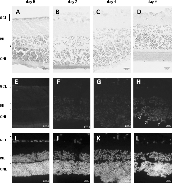
 Figure 5 of
Buonfiglio, Mol Vis 2014; 20:742-752.
Figure 5 of
Buonfiglio, Mol Vis 2014; 20:742-752.