Figure 3. Cx43 downregulation promotes apoptotic death of vascular cells in the retinal capillary networks. Representative images of
capillary networks showing terminal deoxynucleotidyl transferase-mediated uridine 5′-triphosphate-biotin nick end labeling
(TUNEL)-positive cells in the (A) control rats, (B) diabetic rats, (C) rats intravitreally injected with Cx43 siRNA, and (D) rats intravitreally injected with scrambled siRNA. Corresponding images of the 4',6-diamidino-2-phenylindole dihydrochloride
(DAPI)-stained capillary network in (I–L), respectively. The area bound by the dotted line indicates the magnified field of the DAPI-stained capillary network with
a superimposed image from the TUNEL assay (E–H). An increased number of TUNEL-positive cells was present in the retinal capillary networks of the diabetic and Cx43 siRNA-injected
eyes compared with the control groups. M: The graph presents cumulative data showing an increased number of TUNEL-positive cells in the retinal capillaries of the
diabetic rats and the rats intravitreally injected with Cx43 siRNA compared with the control rats. Data are presented as mean
± SD (*p<0.05, n=4). Protein levels of cleaved caspase-3 in the rat retinas of the diabetic rats and those intravitreally
injected with Cx43 siRNA. N: Representative western blot (WB) showing expression of cleaved caspase-3 (upper) in the retinal tissue of normal rats, diabetic
rats, and rats intravitreally injected with Cx43 siRNA or scrambled siRNA. β-actin was used as the loading control (lower).
The graph shows the retinal protein levels of cleaved caspase-3 are significantly increased in diabetic and Cx43 siRNA-injected
rats compared to those of controls. * p<0.05. Scale bar = 50 μm.
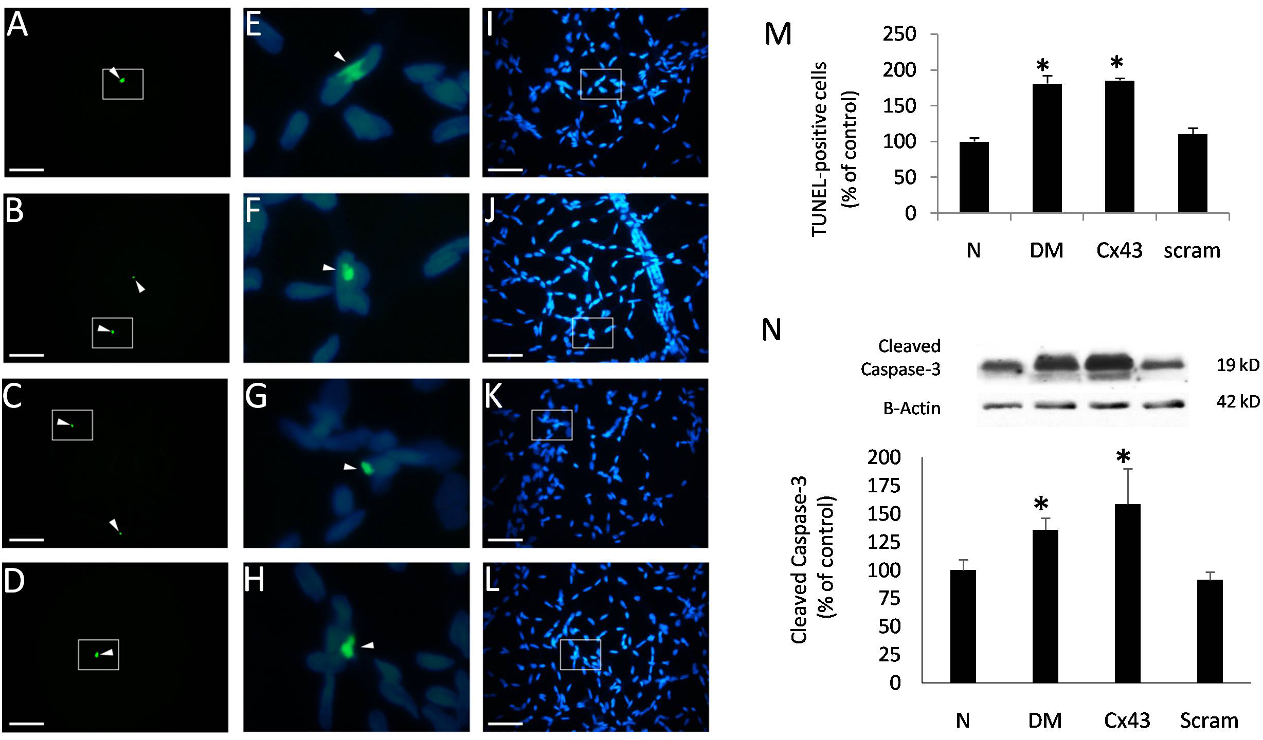
 Figure 3 of
Tien, Mol Vis 2014; 20:732-741.
Figure 3 of
Tien, Mol Vis 2014; 20:732-741.