Figure 2. Cx43 immunostaining rat retinal capillaries. Representative images showing Cx43 immunostaining in the retinal vessels of (A) non-diabetic control rats, (B) diabetic rats, (C) non-diabetic rats intravitreally injected with Cx43 siRNA and (D) non-diabetic rats intravitreally injected with scrambled siRNA with corresponding bright-field images (E–H). Cx43 immunostaining was significantly reduced in the retinal vessels of diabetic rats and those of rats injected with Cx43
siRNA compared with the control rats. Images were captured at 800 ms exposure. Scale bar = 50 μm.
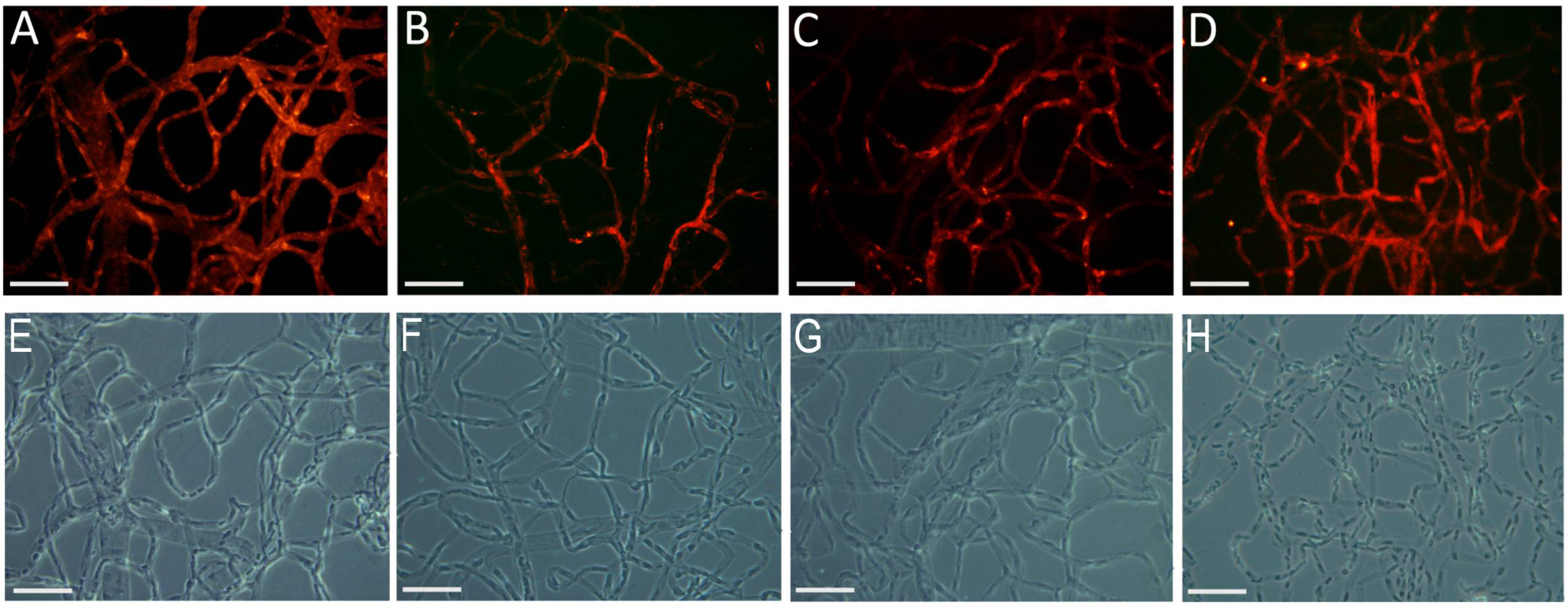
 Figure 2 of
Tien, Mol Vis 2014; 20:732-741.
Figure 2 of
Tien, Mol Vis 2014; 20:732-741.