Figure 1. Cx43 protein levels in diabetic rat retinas and rats intravitreally injected with Cx43 siRNA. A: Representative western blot (WB) showing the Cx43 protein level in the retinas of the control rats, diabetic rats, and rats
treated with intravitreal injections of Cx43 siRNA or scrambled siRNA. Corresponding β-actin protein levels show an equal
amount of protein loaded in each lane (bottom). B: The graph shows retinal Cx43 protein levels are significantly reduced in the diabetic and Cx43 siRNA-injected rats compared
to those of the controls. Data are presented as mean ± SD (* p<0.05, n=4).
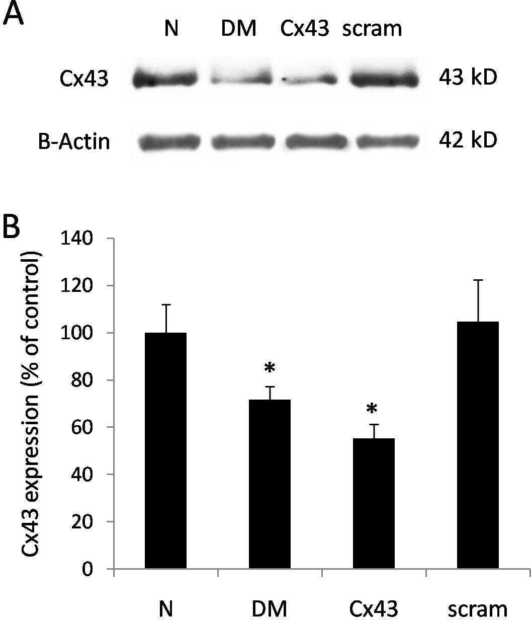
 Figure 1 of
Tien, Mol Vis 2014; 20:732-741.
Figure 1 of
Tien, Mol Vis 2014; 20:732-741.