Figure 3. Fundus fluorescein angiography of vasoproliferative tumor. FFA shows rapid filling of the dye in the early phase, with the
lesion becoming increasingly hyperfluorescent, and profuse and diffuse leakage of dye in the late phase of angiogram.
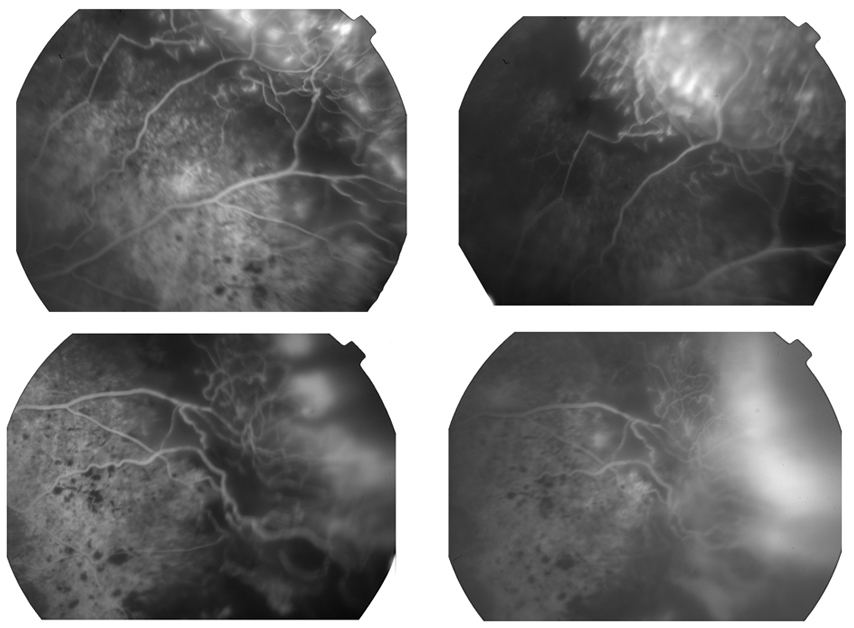
 Figure 3 of
Manayath, Mol Vis 2014; 20:724-731.
Figure 3 of
Manayath, Mol Vis 2014; 20:724-731.