Figure 2. Pre and Post treatment LE color fundus photograph of the proband A: Pre-treatment montage fundus photograph of left eye. LE Fundus shows a healthy appearing optic nerve head with diffuse RPE
degeneration with nummular pigment clumps and white dentritic peripheral vessels in mid peripheral region . Vascularized tumor
mass with exudation and absent feeder vessels is seen in the supro-temporal periphery. B: Vascularized peripheral tumor with profuse exudation. C: 9 Up fundus photograph of LE showing vasoproliferative tumors with exudation in superior, suprotemporal, inferotemporal
and inferior quadrants. D: Post-treatment fundus photograph. LE fundus showing complete regression of the tumor masses with treatment. (6 month post
treatment).
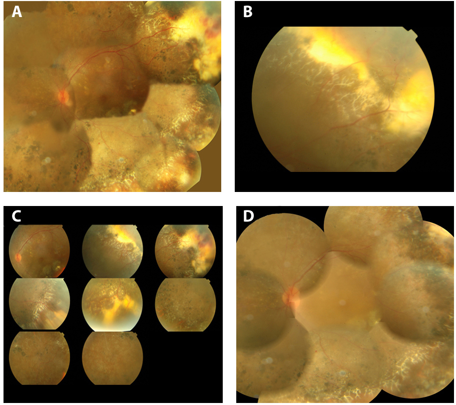
 Figure 2 of
Manayath, Mol Vis 2014; 20:724-731.
Figure 2 of
Manayath, Mol Vis 2014; 20:724-731.