Figure 8. Nitrotyrosine-labeled cells were ionized calcium-binding adaptor molecule-1-positive macrophages in light-damaged choroid.
A–C: Immunolabeling for nitrotyrosine (A), ionized calcium-binding adaptor molecule-1 (Iba-1) (B), and combined images (C), seen here at 6 h post-injury. D–F: Immunolabeling for nitrotyrosine (D), Iba-1 (E), and combined images (F), seen here at 24 h post-injury. Nitrotyrosine was colocalized with Iba-1 expression on macrophages (arrow heads in C and F). Abbreviations: NT, nitrotyrosine; LD, light damage. Scale bar = 50 µm.
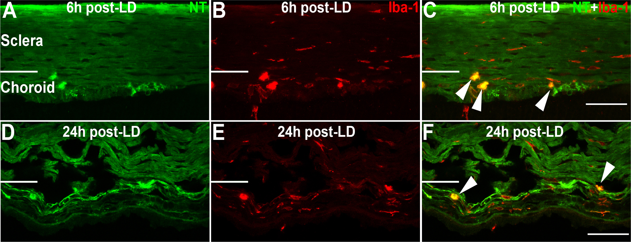
 Figure 8 of
Guo, Mol Vis 2014; 20:670-682.
Figure 8 of
Guo, Mol Vis 2014; 20:670-682.