Figure 7. Colocalization of Cx43 and nitrotyrosine was seen only in the light-damaged choroid. A–C: Cx43 labeling (A), nitrotyrosine labeling (B), and combined images (C) in the non-light-damaged choroid. Immunoreactivity to nitrotyrosine, an oxidative stress marker, was barely detectable.
D–F: Cx43 labeling (D), nitrotyrosine (E), and combined images (F) in the light-damaged choroid at 24 h post-LD. Nitrotyrosine immune-labeled cells showed coexpression of Cx43 (arrow in F) in the choroid. Abbreviations: NT, nitrotyrosine; LD, light damage. Scale bar = 10 µm.
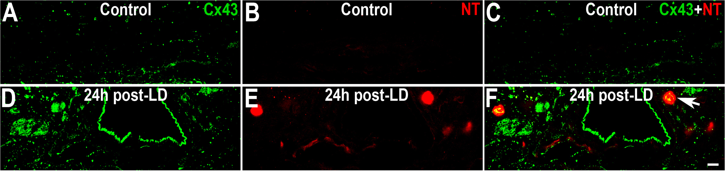
 Figure 7 of
Guo, Mol Vis 2014; 20:670-682.
Figure 7 of
Guo, Mol Vis 2014; 20:670-682.