Figure 4. Cx43 immunoreactivity in the control and light-damaged retinal pigment epithelium and choroid. A: Cx43 immunoreactivity in non-light-damaged control RPE/choroid tissue. Cx43 immunolabeling was seen in the choroid, the
sclera and strongly in the RPE. B–F: Increased Cx43 immunoreactivity was seen in the RPE and choroid tissue from the light-damaged group at the various recovery
time points. Autofluorescence form elastic lamina surrounding a blood vessel is seen in A (arrow). Abbreviations: RPE, retinal pigment epithelium; LD, light damage; BV, blood vessel. Scale bar = 50 µm.
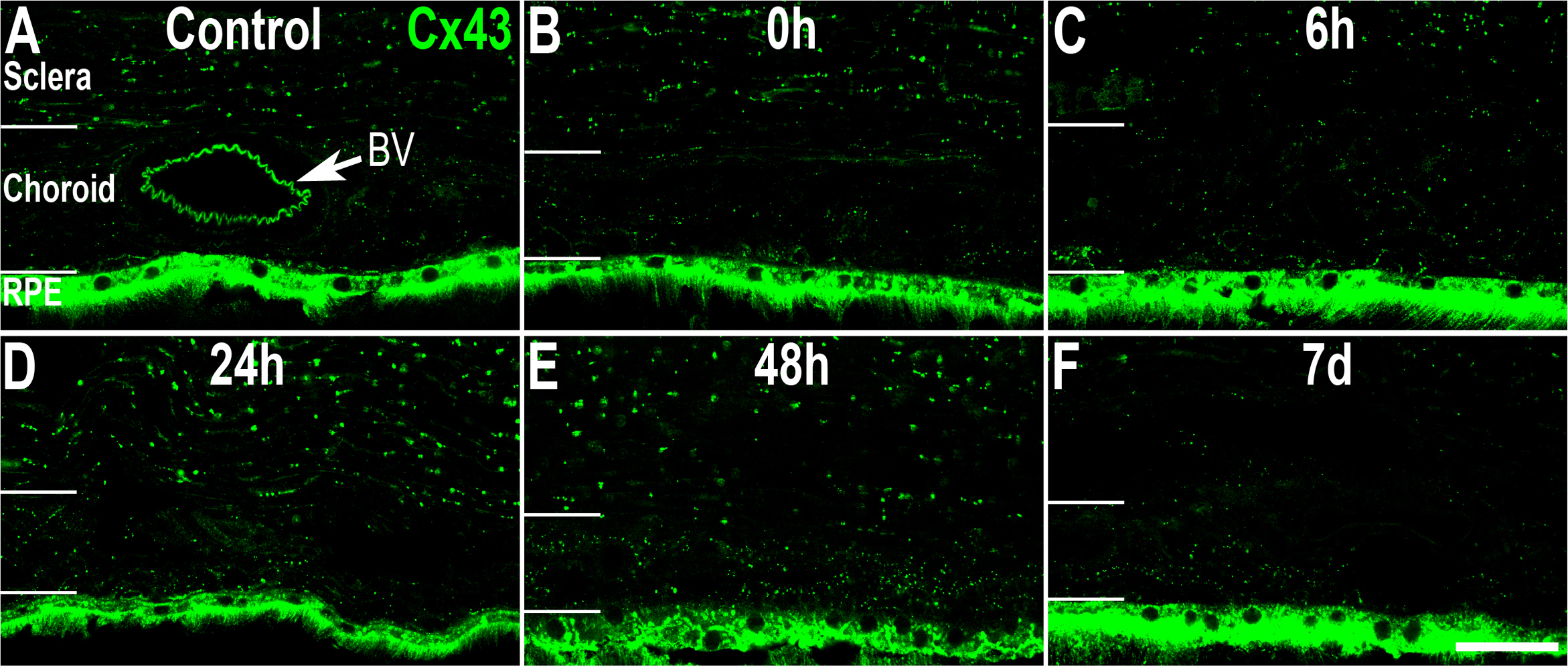
 Figure 4 of
Guo, Mol Vis 2014; 20:670-682.
Figure 4 of
Guo, Mol Vis 2014; 20:670-682.