Figure 3. Western blot analysis of Cx43 expression in the choroid and retina of the control and light-damaged animals. A: Western blot detection of Cx43 in the control and light-damaged rat choroid and RPE. B: Quantitative analysis of western blot for Cx43 protein expression in the choroid and the RPE from the control and light-damaged
groups. Cx43 expression was significantly increased 6 h post-exposure compared to the control (n = 6; ** p<0.01). There was
no statistically significant difference between the control and 0 h groups. C: Western blot detection of Cx43 in the control and light-damaged rat retina. D: Quantitative analysis of western blot for Cx43 expression in the retina from the control and light-damaged groups. The expression
of Cx43 tended to be decreased post-exposure, but this change was not significant (n = 6). Abbreviation: RPE, retinal pigment
epithelium.
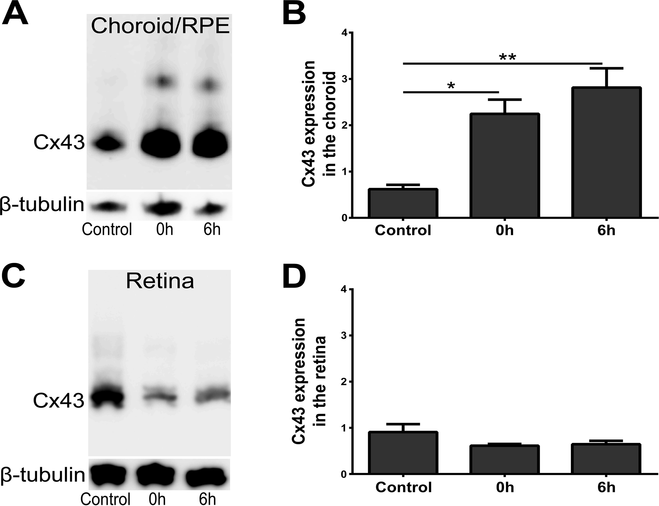
 Figure 3 of
Guo, Mol Vis 2014; 20:670-682.
Figure 3 of
Guo, Mol Vis 2014; 20:670-682.