Figure 2. Cx36 and Cx45 expression in control and light-damaged retina. A: Cx36 showed little immunoreactivity in the retina of non-light-damaged control animals. B: Increased Cx36 expression in the retina collected at 24 h following light damage (LD). C: Cx45 immunoreactivity in the control retina. D: Decreased Cx45 expression in the retina at 24 h following light damage. E–G: Cx36 immunoreactivity in the inner retina from the control rat (E) and light-damaged rat at 24 h (F) and 7 days (G) post-LD. The Cx36 immunoreactivity levels are comparable between the control and 7 days post-LD (n = 5; p = 0.6), but were
significantly increased at 24 h post-LD (see quantification in K, n = 6, *** p<0.001). H–J: Cx45 immunoreactivity in the control retina (H), and the retina at 24 h (I) and 7 days (J) post-LD. The expression levels of Cx45 in the inner plexiform layer were similar and statistically insignificant (n = 6,
p = 0.1) between the control retina and 7 days post-LD. However, there was a marked decrease in Cx45 in the plexiform layer
at the 24 h post injury (I and quantification in L). The control tissue showed stronger Cx45 labeling in the vascular smooth muscle and possibly astrocytes in the nerve fiber
layer (H, arrows), which remained constant at 24 h post-LD (I), but returned to the level comparable to the control by 7 days post-LD (J). All data presented are the mean±standard error of the mean (SEM). Abbreviations: INL, inner nuclear layer; IPL, inner plexiform
layer; GCL, ganglion cell layer; LD, light damage. Scale bar = 50 µm in A–D; scale bar = 20 µm in E–J.
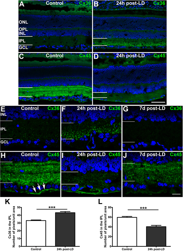
 Figure 2 of
Guo, Mol Vis 2014; 20:670-682.
Figure 2 of
Guo, Mol Vis 2014; 20:670-682.