Figure 2. Cell culture and immunohistochemical staining of malignant melanoma cells. The cells appeared with attached characteristics;
they were spindle-shaped and rich in melanin granules. A: Depigmented cells during the course of culturing. B: Cells passaging to the fifth generation. The melanin granules disappeared completely. C: Illustrates the negative control for immunocytochemical staining. D: Weak positive expression of HMB-45. E: Weak positive expression of Melan-A . F: Positive expression of S-100 (magnification: 200×).
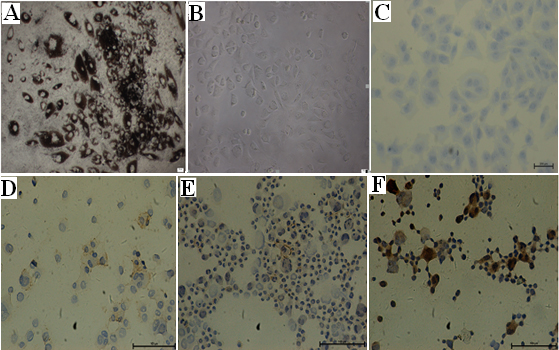
 Figure 2 of
Li, Mol Vis 2014; 20:649-660.
Figure 2 of
Li, Mol Vis 2014; 20:649-660.