Figure 1. An overview, hematoxylin
and eosin (H&E) staining, and immunohistochemical staining
of malignant melanoma. A: An overview of the tumor. B:
H&E staining of the tissue of malignant melanoma. C:
H&E staining depigmented by KMnO4 (potassium
permanganate) of malignant melanoma. The tissue was constituted
by fusiform and epithelioid malignant melanoma cells. Cytoplasm
was abundant with melanin granules. Nuclei was big with
prominent nucleoli. D: Expression of HMB-45 in
immunohistochemical staining of malignant melanoma was positive.
E: Expression of Melan-A was weakly positive; F:
Expression of S-100 protein was positive. G: Negative
control. DAB was used as chromogen. (magnification: 200×).
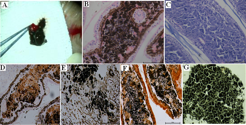
 Figure 1
of Li, Mol Vis 2014; 20:649-660.
Figure 1
of Li, Mol Vis 2014; 20:649-660.