Figure 7. Schematic model of the collagen VI assembly found in human trabecular sheets. N and C termini are depicted in blue and yellow,
respectively, and the collagenous triple helices in red. A: Model for the double-banded aggregates arising from the alignment of the N- and C-globular domains of the collagen VI tetramers.
The black rectangles highlight the position of the transverse bands as seen in the electron micrographs. B: Model for the assemblies presenting a single transverse band. The single band arises from a double band with extra material
(in green) filling the spaces between globular domains.
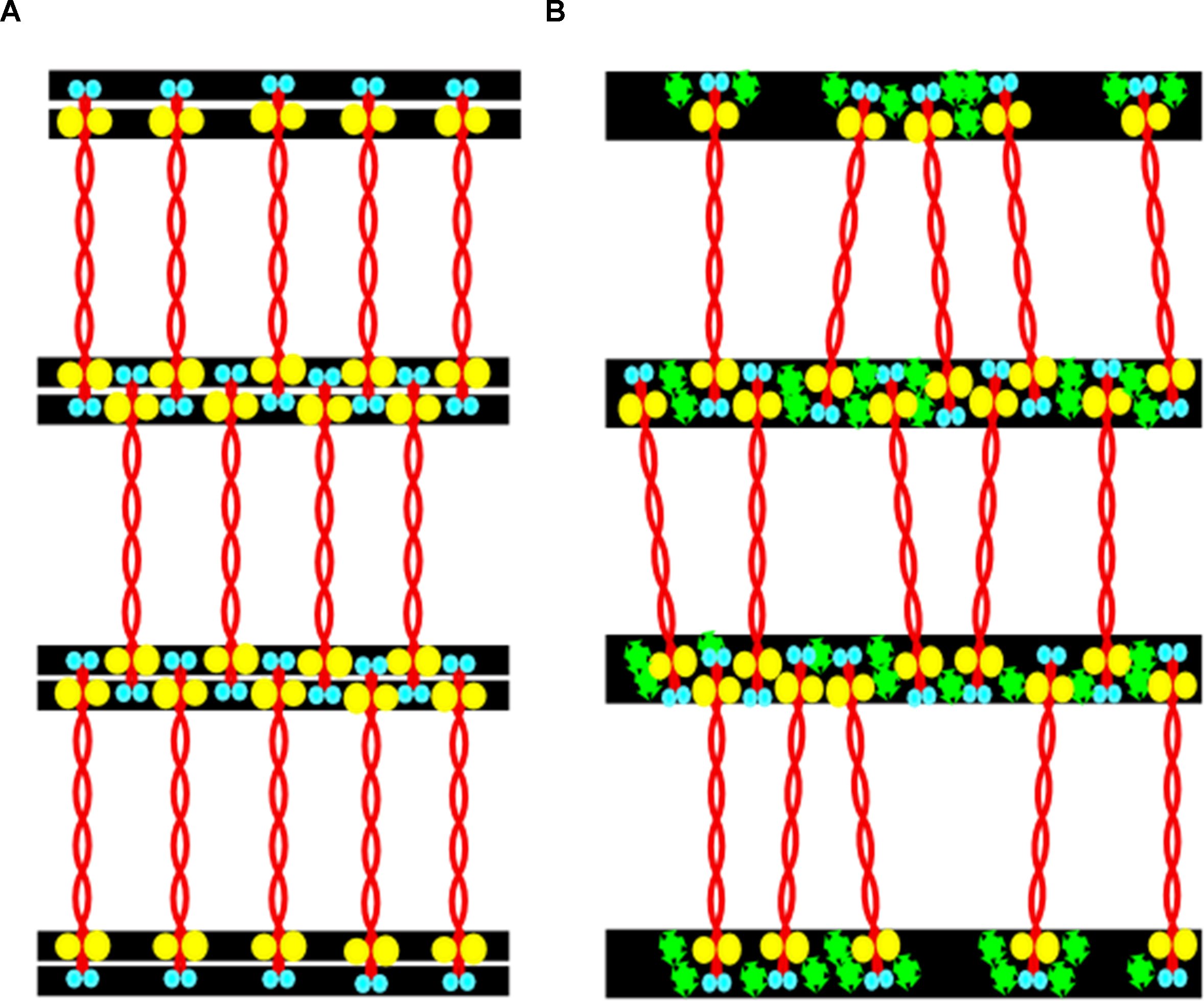
 Figure 7 of
Koudouna, Mol Vis 2014; 20:638-648.
Figure 7 of
Koudouna, Mol Vis 2014; 20:638-648.