Figure 6. Tomographic reconstructions and segmentation of type VI collagen in the human trabecular meshwork. A: An 8.57-nm thick slice through a tomographic reconstruction of a collagen VI assembly. Arrowheads indicate proteoglycans
interacting with collagen VI. B: Two-dimensional surface representation of a three-dimensional (3D) reconstruction. See Appendix 2 for a 360° view of the
reconstruction. The 3D reconstruction has been contrast inverted, and therefore the proteins are shown in white whereas the
background is shown in black. The red arrowheads highlight two globular domains. The tetramer's rod-like segments (yellow
arrowheads) run across the transverse bands and adopt an irregular organization. C: Manually segmented volume of the collagen VI assembly. See Appendix 5 for a 360° view. The transverse bands are shown in
blue, the rod-like segments of the collagen VI tetramers in pink, and proteoglycans in yellow. Proteoglycans vary in size
and interact with the collagen Vl globular domains and the tetramer's rod-like segments. Scale bar = 100 nm.
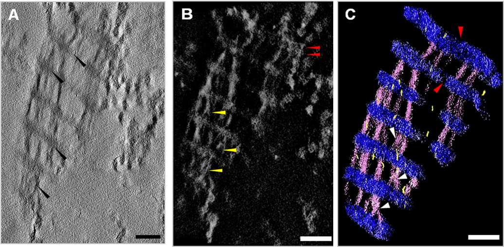
 Figure 6 of
Koudouna, Mol Vis 2014; 20:638-648.
Figure 6 of
Koudouna, Mol Vis 2014; 20:638-648.