Figure 5. High-magnification longitudinal view of the type VI collagen aggregates in the trabecular sheets. Only single transverse bands
are evident (chevrons), and their axial periodicity is approximately 109 nm. The rod-like segments of the collagen VI tetramers
(arrows) cross the transverse bands at different angles. Proteoglycans are also seen in the matrix (arrowheads). Several 10-nm
gold particles used for tomographic reconstruction appear as black dots in the electron micrograph. Scale bar = 200 nm.
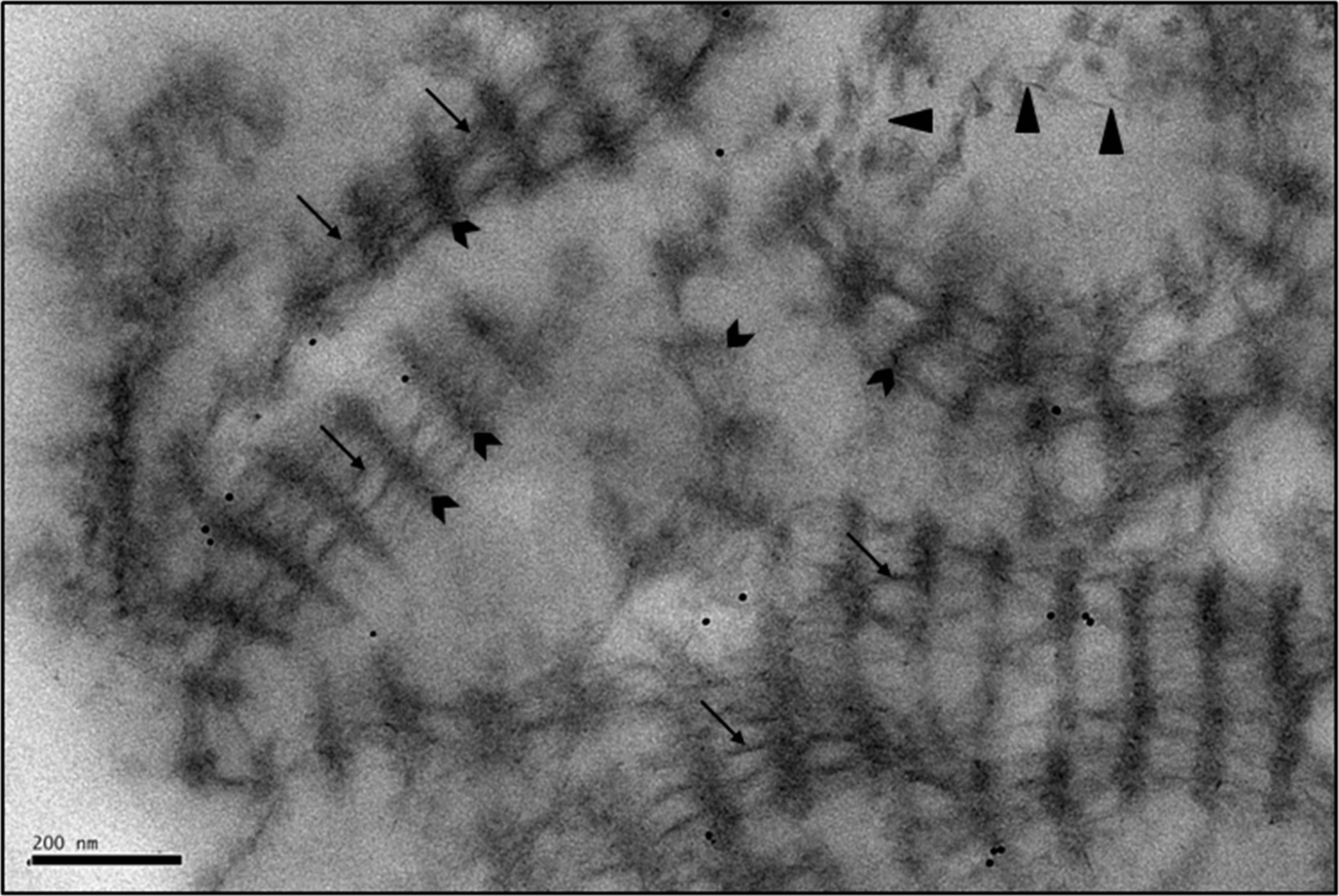
 Figure 5 of
Koudouna, Mol Vis 2014; 20:638-648.
Figure 5 of
Koudouna, Mol Vis 2014; 20:638-648.