Figure 3. Transmission electron micrograph of the human trabecular meshwork showing a series of parallel layers of connective tissue,
known as the trabecular sheets (stars), and intertrabecular spaces forming irregular channels (arrowheads). The trabecular
sheets consist of collagen fibrils orientated both transversely and longitudinally and thin trabecular endothelial cells.
Type VI aggregates (arrows) are distributed throughout the entire width of the sheet surrounded by collagen fibrils. The black
dots dispersed throughout the trabecular meshwork represent 10-nm colloidal gold particles used for tomography. Scale bar
= 1 µm.
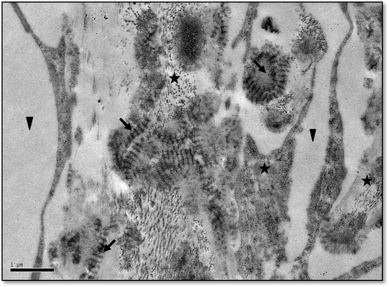
 Figure 3 of
Koudouna, Mol Vis 2014; 20:638-648.
Figure 3 of
Koudouna, Mol Vis 2014; 20:638-648.