Figure 2. Schematic diagram of the eye showing the major ocular structures and the anatomic positions of the Schlemm’s canal (SC), the
juxtacanalicular tissue (JCT), and the trabecular meshwork (TM). The corneoscleral region of the TM is separated from SC by
a connective tissue layer known as the JCT, which terminates adjacent to a layer of endothelial cells that line the inner
wall of SC. The structure of the TM encompasses intertrabecular spaces (IS), trabecular sheets (trabecular beams) filled with
extracellular matrix molecules, including elastin, collagens, some electron-dense dark granules, and thin trabecular endothelial
cells. The IS as well as the trabecular sheets (trabecular beams) vary in size and shape.
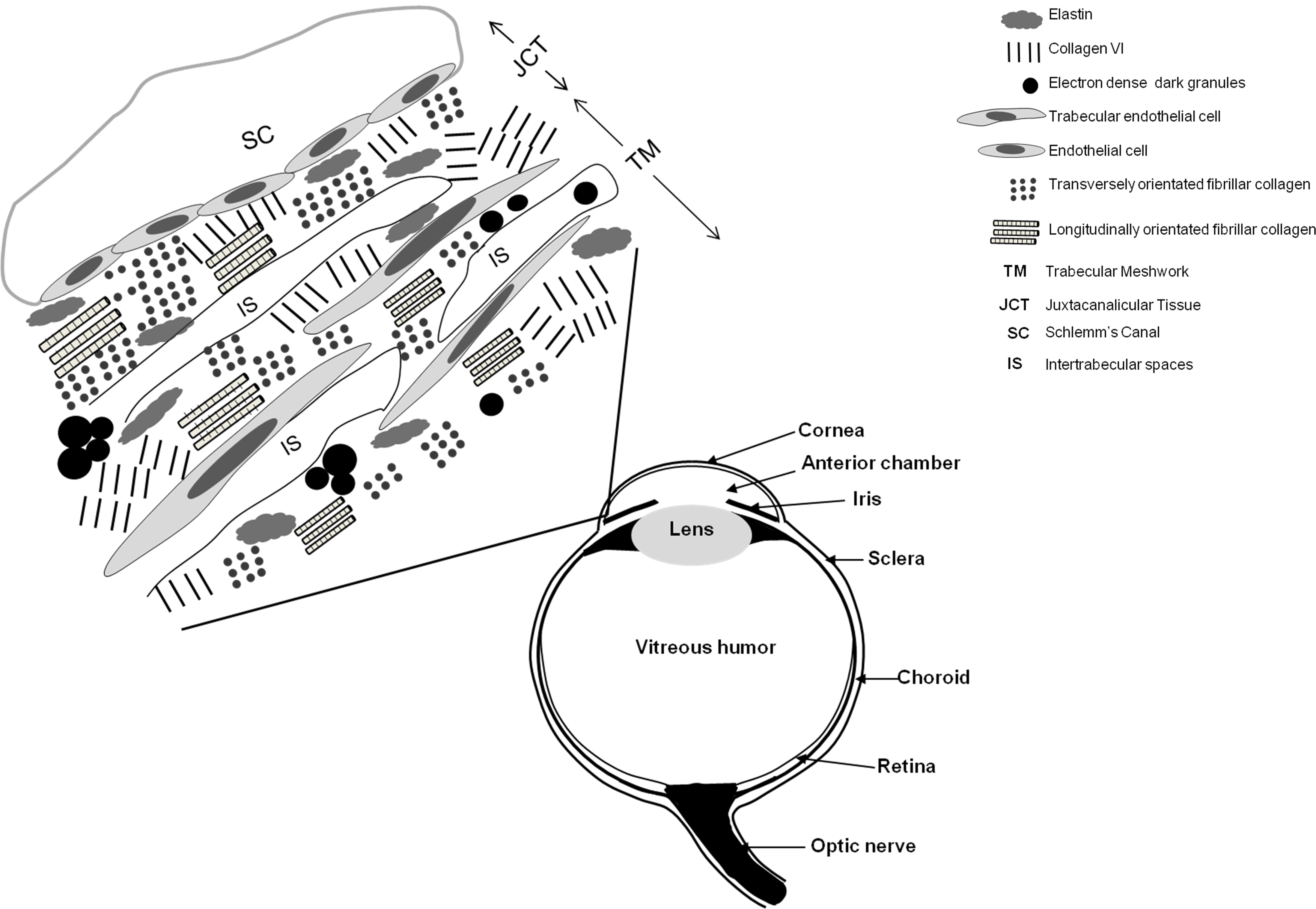
 Figure 2 of
Koudouna, Mol Vis 2014; 20:638-648.
Figure 2 of
Koudouna, Mol Vis 2014; 20:638-648.