Figure 1. Schematic drawings of the structures of (A) monomer, (B) dimer, (C) tetramer, and (D) microfibrils of type VI collagen. N (NH2) and C (COOH) are the amino- and carboxy-terminal domains, represented in blue
and yellow, respectively. The collagenous triple helices (red in color) associate laterally with a 30-nm axial shift in dimers
(B). Pairs of dimers associate laterally to form tetramers (C). Tetramers can be described as possessing an inner rod-like region and two outer rod-like regions separated by and ending
with the collagen VI globular domains. Limited resolution of collagen VI fibrils in micrographs causes the globular domains
to appear as transverse electron-dense bands (D).
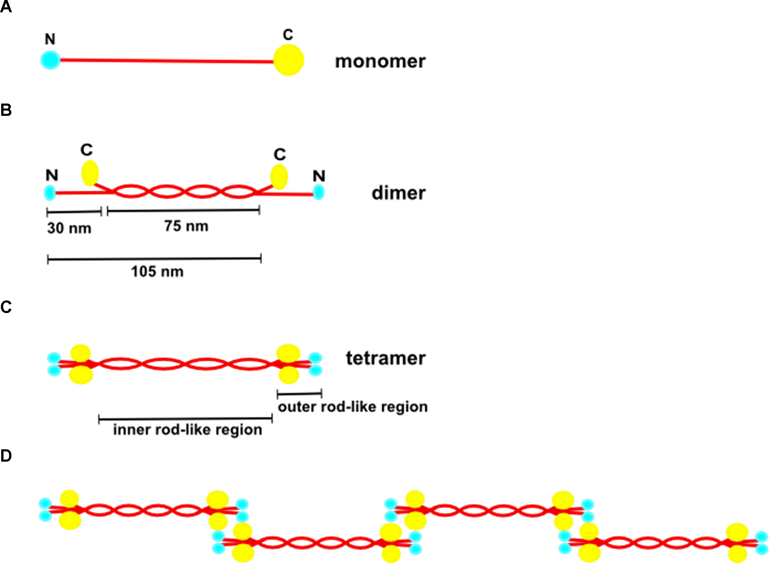
 Figure 1 of
Koudouna, Mol Vis 2014; 20:638-648.
Figure 1 of
Koudouna, Mol Vis 2014; 20:638-648.