Figure 6. Light microscopic examination of the rabbit retina at 3 months (stain, hematoxylin-eosin; magnification, ×400). A: Intravitreal injection of physiologic salt solution. B: Intravitreal injection of 4 mg triamcinolone acetonide (TA). C: Intravitreal injection of 8 mg TA. The three figures show clear retina layers and normal structure. Bar = 20 μm.
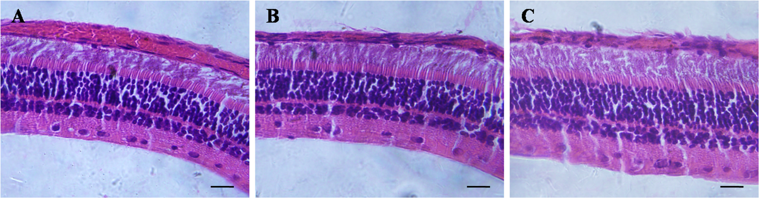
 Figure 6 of
Ye, Mol Vis 2014; 20:629-636.
Figure 6 of
Ye, Mol Vis 2014; 20:629-636.