Figure 4. Tumor necrosis factor-α (TNF-α) was released from the activated microglia induced by retinal neuronal monocyte chemoattractant
protein-1 (MCP-1) via the phosphorylation of extracellular signal-regulated kinase (ERK). A: Real-time PCR was used to measure TNF-α mRNA expression. Dose-dependent inhibition of the expression of TNF-α mRNA was induced
by retinal neuronal MCP-1 in the retinal neuron–microglia Transwell culture system by ERK inhibitors. B: Enzyme-linked immunosorbent assay (ELISA) was used to measure the soluble TNF-α concentration. Dose-dependent inhibition
of the expression of soluble TNF-α was induced by ERK inhibitors. C: Phospho-ERK levels were detected with western blotting. Phospho-ERK levels from the microglial cells increased due to retinal
neuronal MCP-1; however, anti-MCP-1 led to downregulation of the levels of phospho-ERK (**p<0.01). D: Purified microglia were stained with fluorescein isothiocyanate (FITC)-CD11b (green), and the expression of TNF-α was labeled
with PE (red). CD11b and TNF-α double-stained cells were detectable in the control medium. The number of double-stained cells
(activated microglia) increased markedly after the advanced glycation end product (AGE) treatment. In contrast, the number
of CD11b and TNF-α double-stained cells (activated microglia) decreased markedly after AGE treatment with ERK inhibitors (control:
14.58±4.54 cells/microscopic visual field; AGE: 38.84±6.11 cells/microscopic visual field; ERK inhibitors: 26.25±3.66 cells/microscopic
visual field; p=0.021).
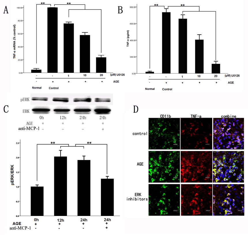
 Figure 4 of
Dong, Mol Vis 2014; 20:616-628.
Figure 4 of
Dong, Mol Vis 2014; 20:616-628.