Figure 2. Effects of retinal neuronal monocyte chemoattractant protein-1 (MCP-1) on activated microglial cells. A: Tumor necrosis factor-α (TNF-α) mRNA from microglia was significantly stimulated by exogenous MCP-1 and CC-chemokine receptor
2 (CCR2) knockdown, which led to downregulation of TNF-α mRNA from microglia. B: The concentrations of soluble TNF-α were significantly stimulated by exogenous MCP-1. In addition, CCR2 knockdown led to
downregulation of TNF-α release from microglia induced by exogenous MCP-1. C: The expression of TNF-α mRNA was significantly increased by neuronal MCP-1 in the retinal neuron–microglia Transwell culture
system. In addition, TNF-α mRNA expression was reduced when the MCP-1 blocking peptide and CCR2 siRNA were used. However,
the incubation of control immunoglobulin G (IgG) or control siRNA did not reduce the TNF-α production. D: Enzyme-linked immunosorbent assay (ELISA) was used to measure the soluble TNF-α concentration in the retinal neuron–microglia
Transwell culture system. When cultured with advanced glycation end products (AGEs), the retinal neurons had a markedly increased
effect on retinal microglial activation in the Transwell culture system. In addition, the concentration of soluble TNF-α was
decreased when the MCP-1 blocking peptide and CCR2 siRNA were used. However, the incubation of control IgG or control siRNA
did not reduce TNF-α production. Data shown are the mean±SD (**p<0.01).
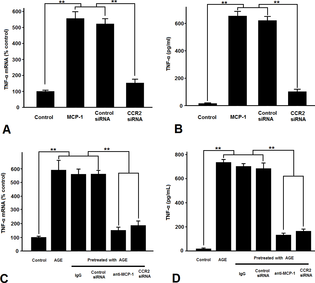
 Figure 2 of
Dong, Mol Vis 2014; 20:616-628.
Figure 2 of
Dong, Mol Vis 2014; 20:616-628.