Figure 6. Effect of phosphatidylinositol 3-kinase inhibitor on wound healing after gene silencing (n = 4). The scratch wound was created
using 1 ml sterile serological pipette in a confluent corneal fibroblast culture after gene silencing of β-actin or human
antigen R (HuR). The images were taken at 0 h and 24 h. The lines show the area where the scratch wound was created. A: Scratch wound at 0 h in cells that were treated with scrambled siRNA. B: Migration of cells to the site of the wound after 24 h in cells that was treated with scrambled siRNA. C: Migration of cells to the site of the wound after 24 h in cells that were treated with scrambled siRNA + phosphatidylinositol
3-kinase (PI3K) inhibitor Ly292004. D: A scratch wound at 0 h in cells that were treated with β-actin siRNA. E: Migration of cells to the site of the wound after 24 h that were treated with β-actin siRNA. F: Migration of cells to the site of wound after 24 h in cells that was treated with β-actin siRNA +PI3 kinase inhibitor Ly292004.
G: A scratch wound at 0 h in cells that were treated with HuR siRNA. H: Migration of cells to the site of the wound after 24 h in cells that were treated with HuR siRNA. I: Migration of cells to the site of the wound after 24 h in cells that was treated with HuR siRNA +PI3 kinase inhibitor Ly292004.
The scratch wound assay was performed in tetraplicate. J: A schematic representation of PI3K-induced actin filament remodeling and migration.
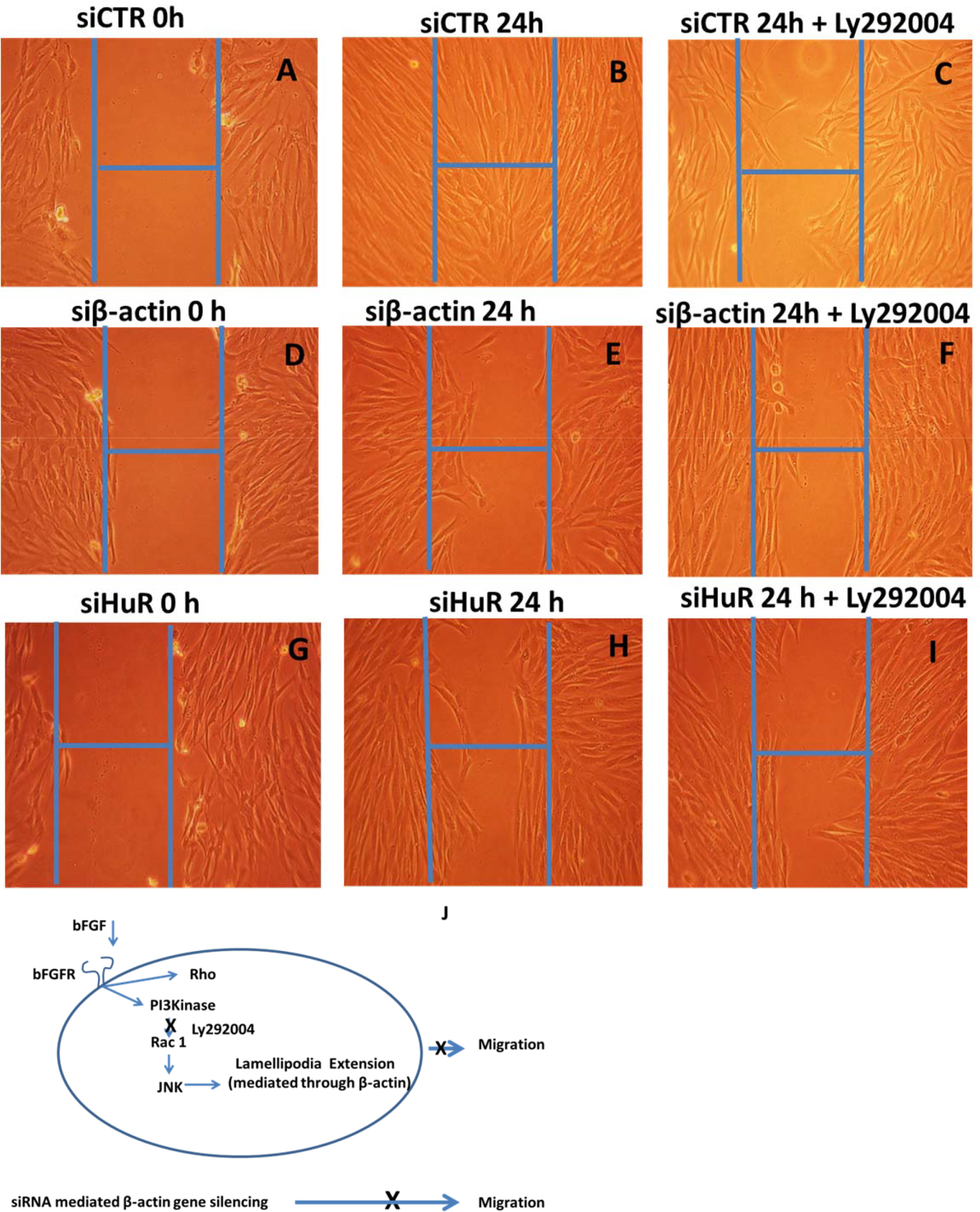
 Figure 6 of
Joseph, Mol Vis 2014; 20:593-605.
Figure 6 of
Joseph, Mol Vis 2014; 20:593-605.