Figure 5. Effect of wound healing after gene silencing (n = 4). A scratch wound was created using 1 ml sterile serological pipette in
a confluent corneal fibroblast culture after gene silencing of GAPDH, β-actin, or human antigen R (HuR). The images were taken
at 0 h and 24 h. The dotted lines show the area where the scratch wound was created. A: Normal cells with a scratch wound at 0 h. B: Cells migrating to the site of wound after 24 h. C: Scratch wound at 0 h in cells that were treated with scrambled siRNA. D: Migration of cells to the site of wound after 24 h in cell culture that was treated with scrambled siRNA. E: Scratch wound at 0 h in cells that were treated with GAPDH siRNA. F: Migration of cells to the site of wound after 24 h that were treated with scrambled siRNA. G: Scratch wound at 0 h in cells that were treated with HuR siRNA. H: Migration of cells to the site of wound after 24 h that were treated with HuR siRNA. I: Scratch wound at 0 h in cells that were treated with β-actin siRNA. J: Cells migrated to the site of wound after 24 h in cells that were treated with β-actin siRNA. K: A bar graph showing the number of cells that migrated after 24 h following the scratch wound in cells treated with siScrambled,
siGAPDH, siHuR, or siβ-actin. The scratch wound assay was performed in tetraplicate, and standard deviations were calculated.
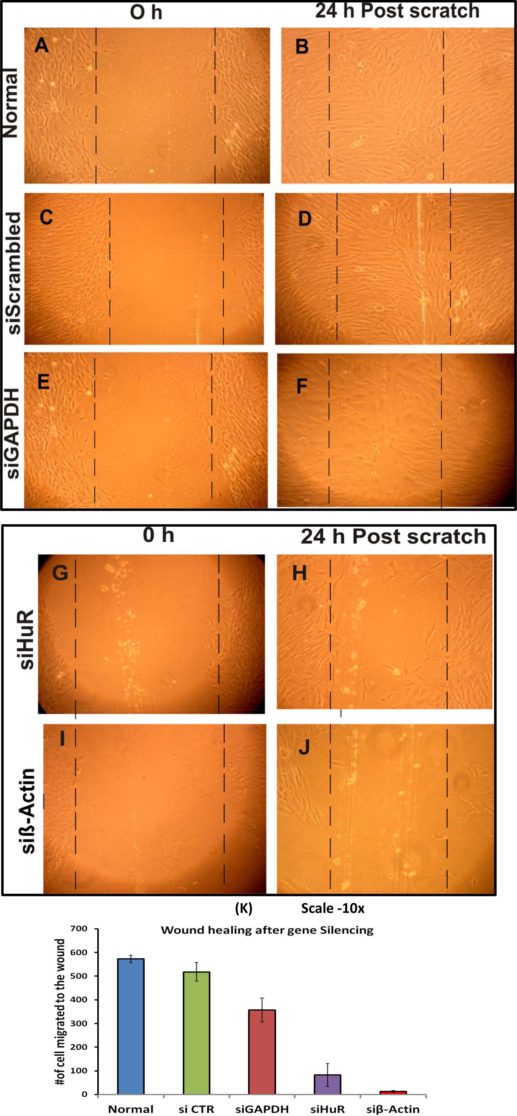
 Figure 5 of
Joseph, Mol Vis 2014; 20:593-605.
Figure 5 of
Joseph, Mol Vis 2014; 20:593-605.