Figure 3. Localization of β-actin in corneal fibroblasts after gene silencing. The normal corneal fibroblasts were treated with scrambled
siRNA, GAPDH siRNA, β-actin siRNA, or human antigen R (HuR) siRNA. The immunoreactivity of anti-β-actin antibody was analyzed
at 72 h after transfection. A: Fibroblasts stained with Hoechst nuclear stain after treatment with scrambled siRNA. B: Immunoreactivity of fibroblasts with anti-β-actin antibody after treatment with scrambled siRNA. C: Fibroblasts stained with rhodamine-labeled phalloidin stain after treatment with scrambled siRNA. D: Overlay of (A), (B), and (C). E: Fibroblasts stained with Hoechst nuclear stain after treatment with GAPDH siRNA. F: Immunoreactivity of fibroblasts with anti-β-actin antibody after treatment with GAPDH siRNA. G: Fibroblasts stained with rhodamine-labeled phalloidin stain after treatment with GAPDH siRNA. H: Overlay of (E), (F), and (G). I: Fibroblasts stained with Hoechst nuclear stain after treatment with HuR siRNA. J: Immunoreactivity of fibroblasts with anti-β-actin antibody after treatment with HuR siRNA. K: Fibroblasts stained with rhodamine-labeled phalloidin stain after treatment with HuR siRNA. L: Overlay of (I), (J), and (K). M: Fibroblasts stained with Hoechst nuclear stain after treatment with β-actin siRNA. N: Immunoreactivity of fibroblasts with anti-β-actin antibody after treatment with β-actin siRNA. O: Fibroblasts stained with rhodamine-labeled phalloidin stain after treatment with β-actin siRNA, and (P): Overlay of (M), (N), and (O). Note that β-actin and HuR gene silencing affected β-actin expression. Scrambled siRNA transfection of normal corneal fibroblasts showed no effect on
β-actin expression, whereas GAPDH siRNA treatment had a drastic effect on β-actin expression.
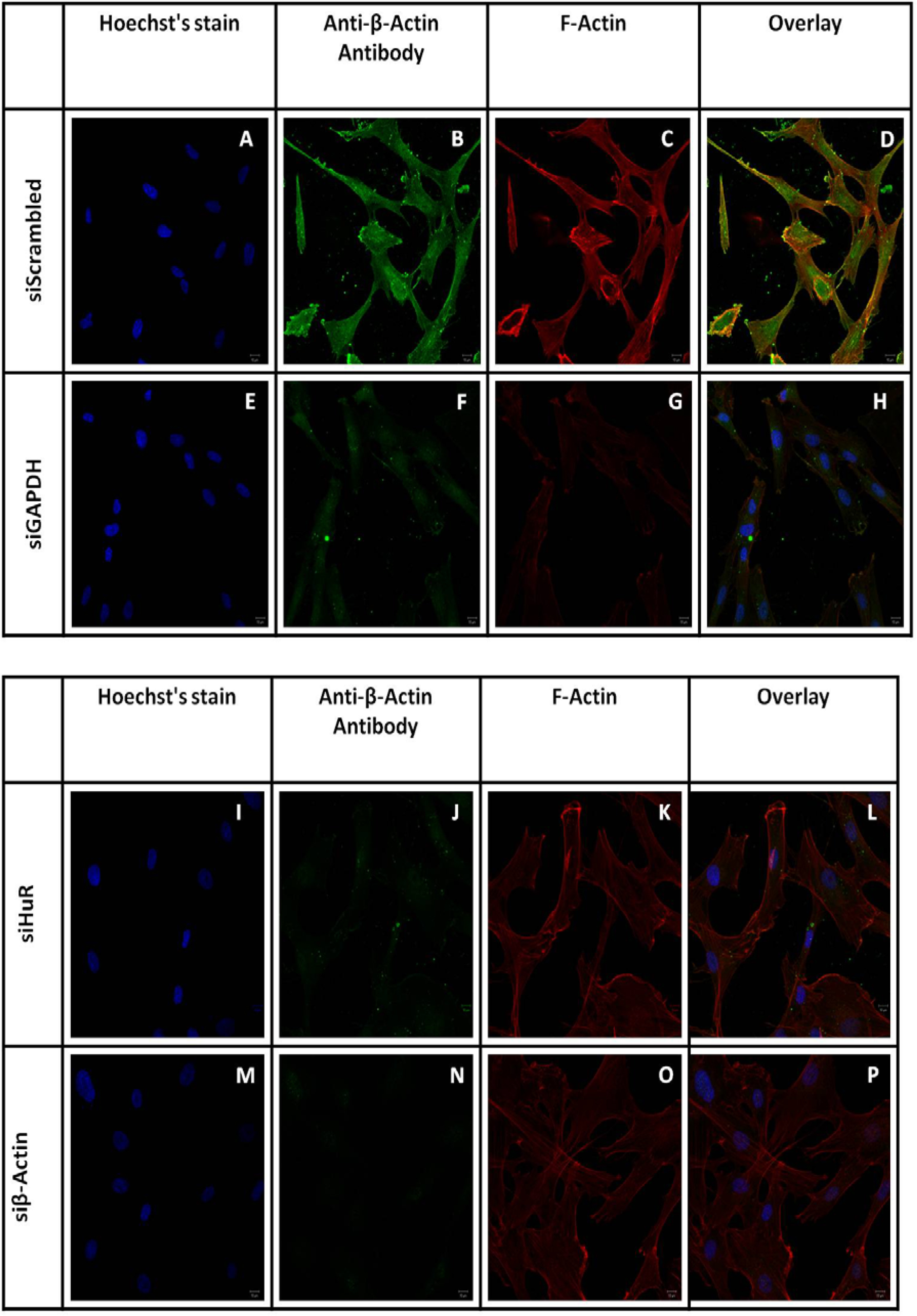
 Figure 3 of
Joseph, Mol Vis 2014; 20:593-605.
Figure 3 of
Joseph, Mol Vis 2014; 20:593-605.