Figure 7. Patient #21.Blue fundus autofluorescence (FAF) and spectral-domain optical coherence tomography (SD-OCT) reveal the right
eye affected with vitelliruptive lesion at both study entry and last follow-up visit (50 months later). Blue FAF frames and
SD-OCT scans at study entry (top left and bottom right panels) show reabsorption of the hyperautofluorescent/hyperreflective
subretinal material (asterisk) and replacement by a fluid component. At the last follow-up visit, blue FAF remained almost
unchanged (top right panel), while SD-OCT showed a decrease in subretinal fluid (asterisk; bottom right panel).
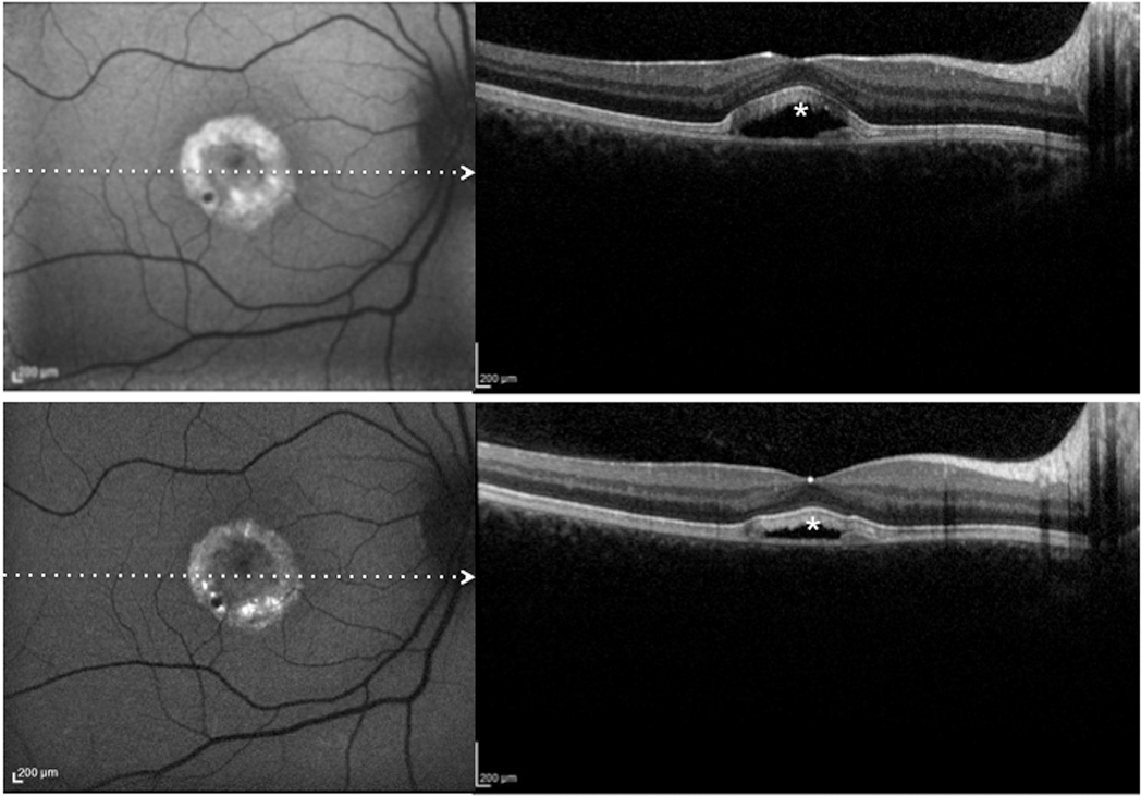
 Figure 7 of
Querques, Mol Vis 2014; 20:575-592.
Figure 7 of
Querques, Mol Vis 2014; 20:575-592.