Figure 6. Patient #1.Blue fundus autofluorescence (FAF) and spectral-domain optical coherence tomography (SD-OCT) reveal the right eye
affected with pseudohypopyon lesion at both study entry and last follow-up visit (61 months later). Blue FAF frames and SD-OCT
scans at study entry (top left and bottom right panels) show a partial reabsorption of the hyperautofluorescent (arrowhead)/hyperreflective
material (asterisk) and replacement by a fluid component. During follow-up, blue FAF frames and SD-OCT scans (top middle and
bottom middle panels) show further reabsorption of the hyperautofluorescent (arrowhead)/hyperreflective material (asterisk).
At the last follow-up visit, blue FAF frames and SD-OCT scans (top left and bottom right panels) show development of the hyperautofluorescent
(arrowhead)/hyper-reflective material (asterisk).
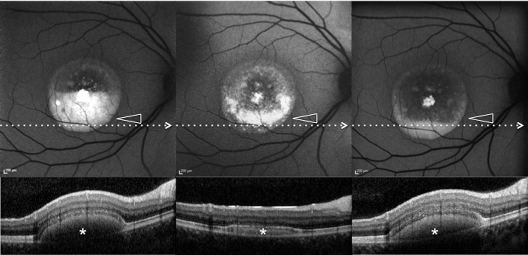
 Figure 6 of
Querques, Mol Vis 2014; 20:575-592.
Figure 6 of
Querques, Mol Vis 2014; 20:575-592.