Figure 8. Interleukin-6 signaling in wild-type and Tmprss6msk/msk mouse retinas. A: Immunofluorescent analysis of interleukin-6 (IL-6) expression in wild-type (WT) and Tmprss6msk/msk mouse retinas using cryosections. The experiment was repeated with two mice (four retinas) for each genotype. B: qPCR analysis of IL-6 mRNA levels in 3-month-old and 5-month-old WT and Tmprss6msk/msk mouse retinas. Data are presented as the fold change in the Tmprss6msk/msk retina relative to the WT retina. Data represent the results from four independent RNA preparations (four wild-type mice
and four Tmprss6msk/msk mice). *, p<0.05; ***, p<0.001. C: Immunofluorescent analysis of the expression of the phosphorylated form of STAT3 (pSTAT3) in wild-type (WT) and Tmprss6msk/msk mouse retinas using cryosections. The experiment was repeated with two mice (four retinas) for each genotype.
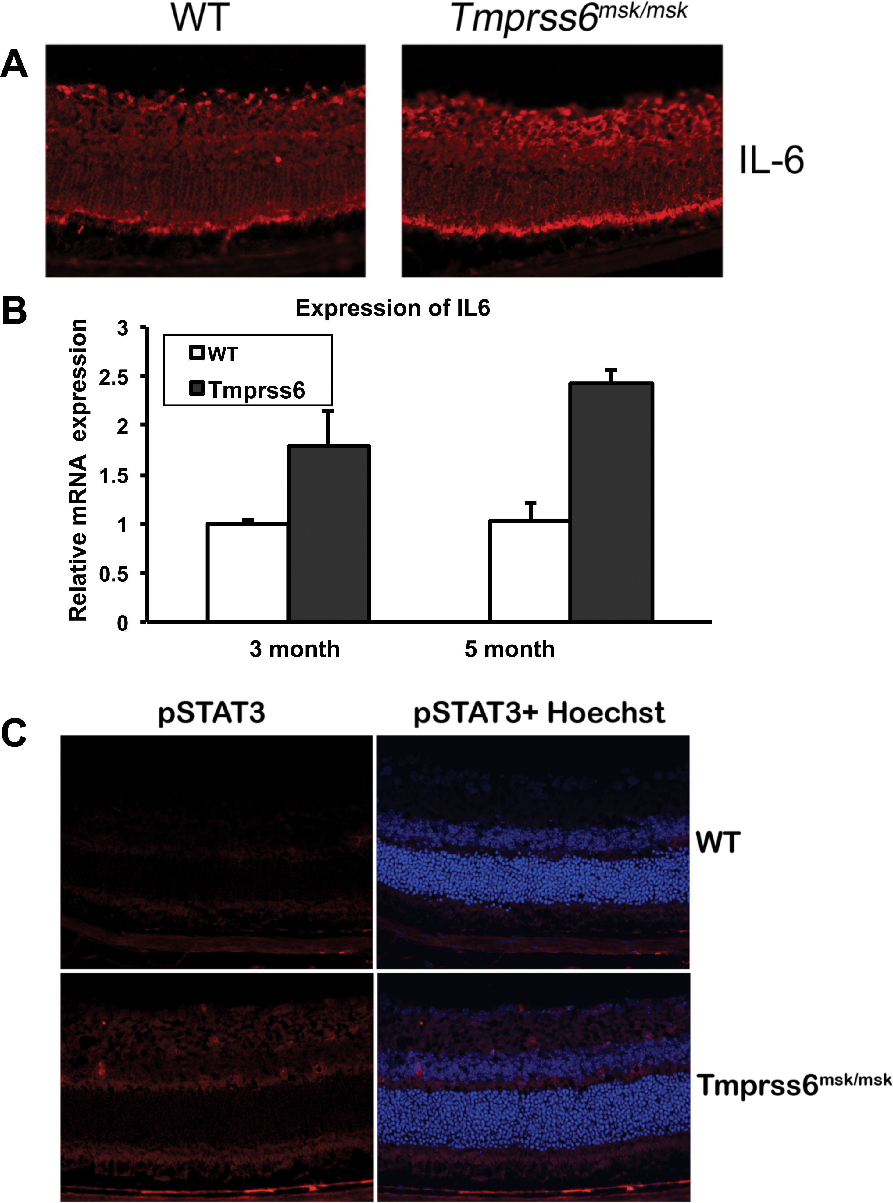
 Figure 8 of
Gnana-Prakasam, Mol Vis 2014; 20:561-574.
Figure 8 of
Gnana-Prakasam, Mol Vis 2014; 20:561-574.