Figure 2. Specificity of matriptase-2 staining in retina and retinal morphology in wild-type and Tmprss6msk/msk mice. A: The expression pattern of matriptase-2 was analyzed in retinal sections prepared from wild-type mice and Tmprss6msk/msk mice using an antibody specific for the C-terminal tail of the matriptase-2 protein. The Tmprss6msk/msk mouse expresses a truncated matriptase-2 that lacks the C-terminal tail. The experiment was repeated twice with retinal sections
from different wild-type mice and Tmprss6msk/msk mice. B: Retinal morphology as assessed with hematoxylin and eosin staining of retinal sections from wild-type and Tmprss6msk/msk mice. Eyes from 5-month-old wild-type and Tmprss6msk/msk mice were enucleated, fixed, and embedded in JB-4. Sections were cut at 2-μm thickness and examined with light microscopy.
The morphology was examined individually with retinal sections prepared from four wild-type mice and four Tmprss6msk/msk mice, and the results were similar.
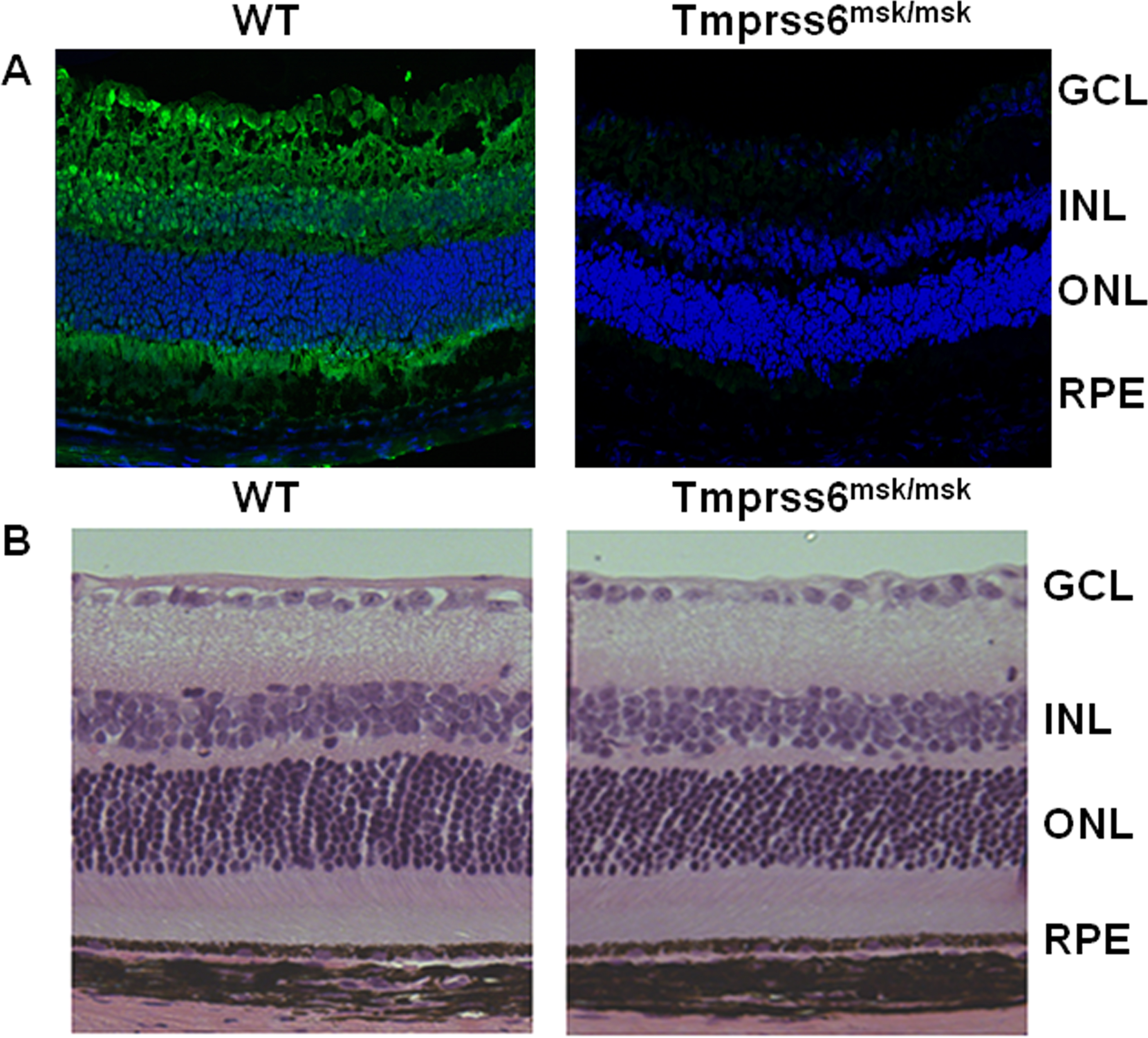
 Figure 2 of
Gnana-Prakasam, Mol Vis 2014; 20:561-574.
Figure 2 of
Gnana-Prakasam, Mol Vis 2014; 20:561-574.