Figure 1. Expression of matriptase-2 in the mouse retina. A: RT–PCR analysis of mRNAs for matriptase-2, neogenin, and hemojuvelin in the neural retina (NR) and the RPE/eyecup and in
primary cultures of RPE (1°RPE), Müller (1°MC), and ganglion (1°GC) cells isolated from mouse eyes. 18S was used as an internal
control. RT–PCR was repeated with RNA preparations from three different mice, and the results were similar. B: Immunofluorescence localization of matriptase-2 protein in the mouse retina: left-hand panel, nuclear specific Hoechst staining;
right-hand panel, pattern of matriptase-2 expression. GCL, ganglion cell layer; INL, inner nuclear layer; ONL, outer nuclear
layer; RPE, retinal pigment epithelium. C: Polarized expression of matriptase-2 in RPE investigated by comparing its expression pattern with that of monocarboxylate
transporter 1 (MCT1; a marker of RPE apical membrane) and Hfe (a marker for basolateral membrane). In addition, the relationship
between the expression pattern in the RPE of matriptase-2 with that of neogenin (a protein that interacts with hemojuvelin)
and hemojuvelin (the substrate for matriptase-2) was also studied. The immunocomplexes were detected with appropriate secondary
antibodies conjugated to either Alexa Fluor 568 (red) or Alexa Fluor 488 (green). Immunofluorescence localization studies
were repeated with retinal sections prepared from two different mice.
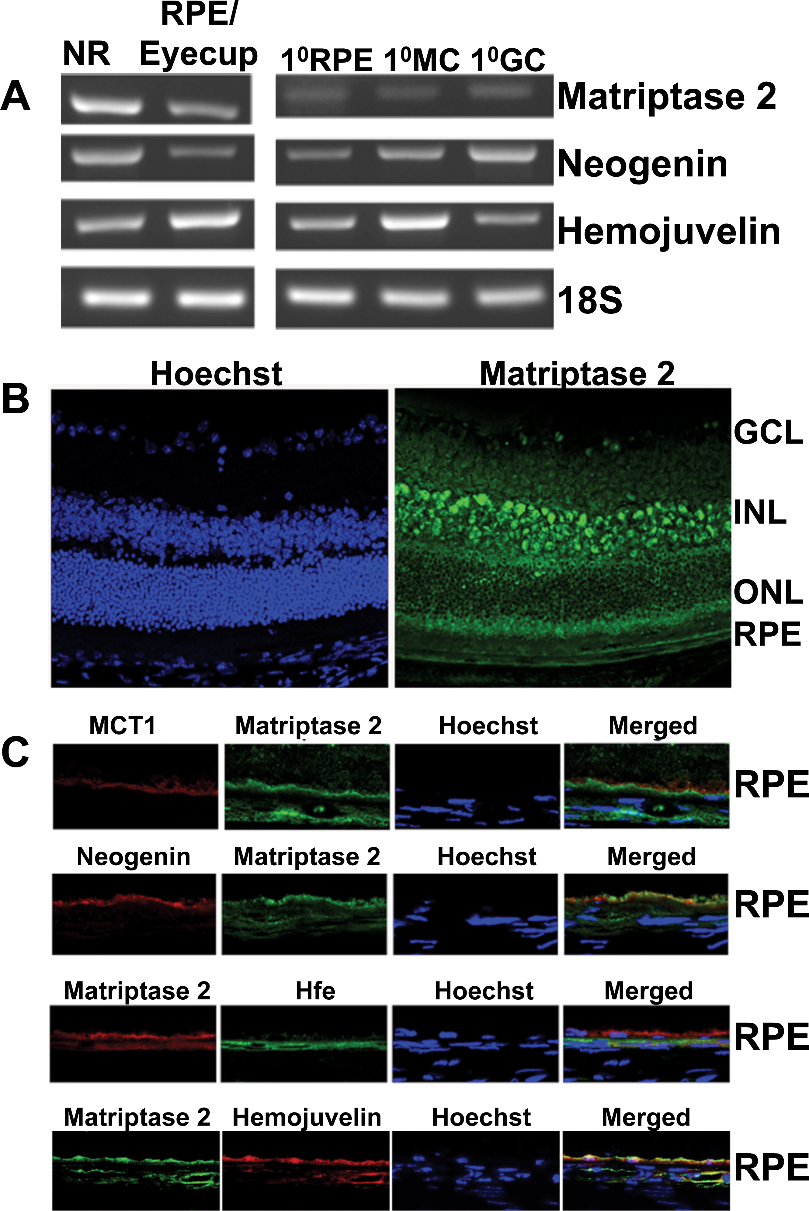
 Figure 1 of
Gnana-Prakasam, Mol Vis 2014; 20:561-574.
Figure 1 of
Gnana-Prakasam, Mol Vis 2014; 20:561-574.