Figure 2. Bilateral fundus photography of the probands from the respective families with retinal dystrophies. Bilateral fundus photography
shows attenuation of retinal arterioles, atrophy of the retinal pigment epithelium, and the waxy-pale disc of both eyes in
all fundus photograph. Bone spicule-like pigmentation was found in the periphery retina of patient III:4 in Family-012, sheet
pigmentation was found in the posterior pole retina of patient II:6 in Family-024, and macular coloboma was found in patient
II:2 in Family-035.
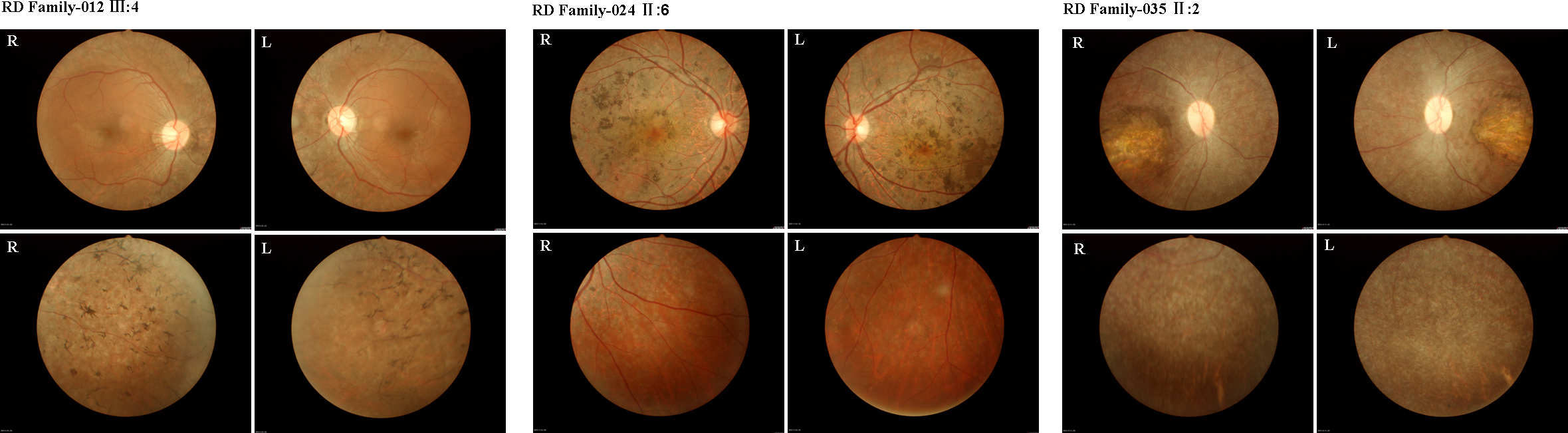
 Figure 2 of
Jin, Mol Vis 2014; 20:553-560.
Figure 2 of
Jin, Mol Vis 2014; 20:553-560.