Figure 1. The effects of transforming growth factor-beta on mesenchymal marker and limbal epithelial stem cell marker expression in
cultured limbal epithelial stem cells. A: Dosage effect on P1 limbal epithelial stem cells (LSCs). P1 cells were treated with 2–20 ng/mL transforming growth factor-beta
(TGFβ1) for 48 h, and then processed for western blot analysis. B: P0 and P1 LSCs were treated with 10 ng/ml TGFβ1 for 48 h, and proteins were detected with western blot analysis with antibodies
as indicated. Representative blots (A and B) and densitometric analysis with standard deviation (SD; C) of four independent experiments are shown. Untreated P0 cells (UT) set as onefold or 100%. *p<0.005 versus TGFβ1-treated
P1 cells. **p<0.01 versus TGFβ1-treated P1 cells.
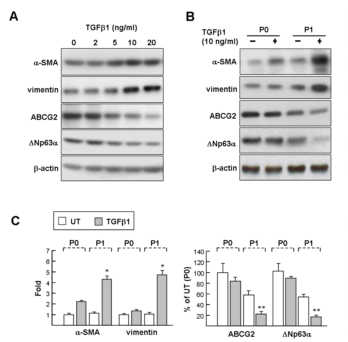
 Figure 1 of
Tsai, Mol Vis 2014; 20:522-534.
Figure 1 of
Tsai, Mol Vis 2014; 20:522-534.