Figure 5. Picrosirius red (left) and negative control–Mayer’s hematoxylin only (right) stained sagittal sections of paraffin-embedded
basset hound (BH) eyes. Images were viewed using polarizing light microscopy. Collagen staining of fibers appears in hues
of orange, pink, and red. The observed color intensity is indicative of collagen fiber thickness and abundance in tissue.
Collagen fiber staining was noted in A: the ciliary body (CB) and processes, B: stroma and pigmentary epithelium (PE) layers of the iris, C: epithelium (EP), stroma and endothelium (ED) layers of the cornea, D: optic nerve (ON), E: ciliary cleft (CC) and trabecular meshwork (TM). Arrows indicate areas where staining was prominently observed, including
the CB, iris, lens, and ON.
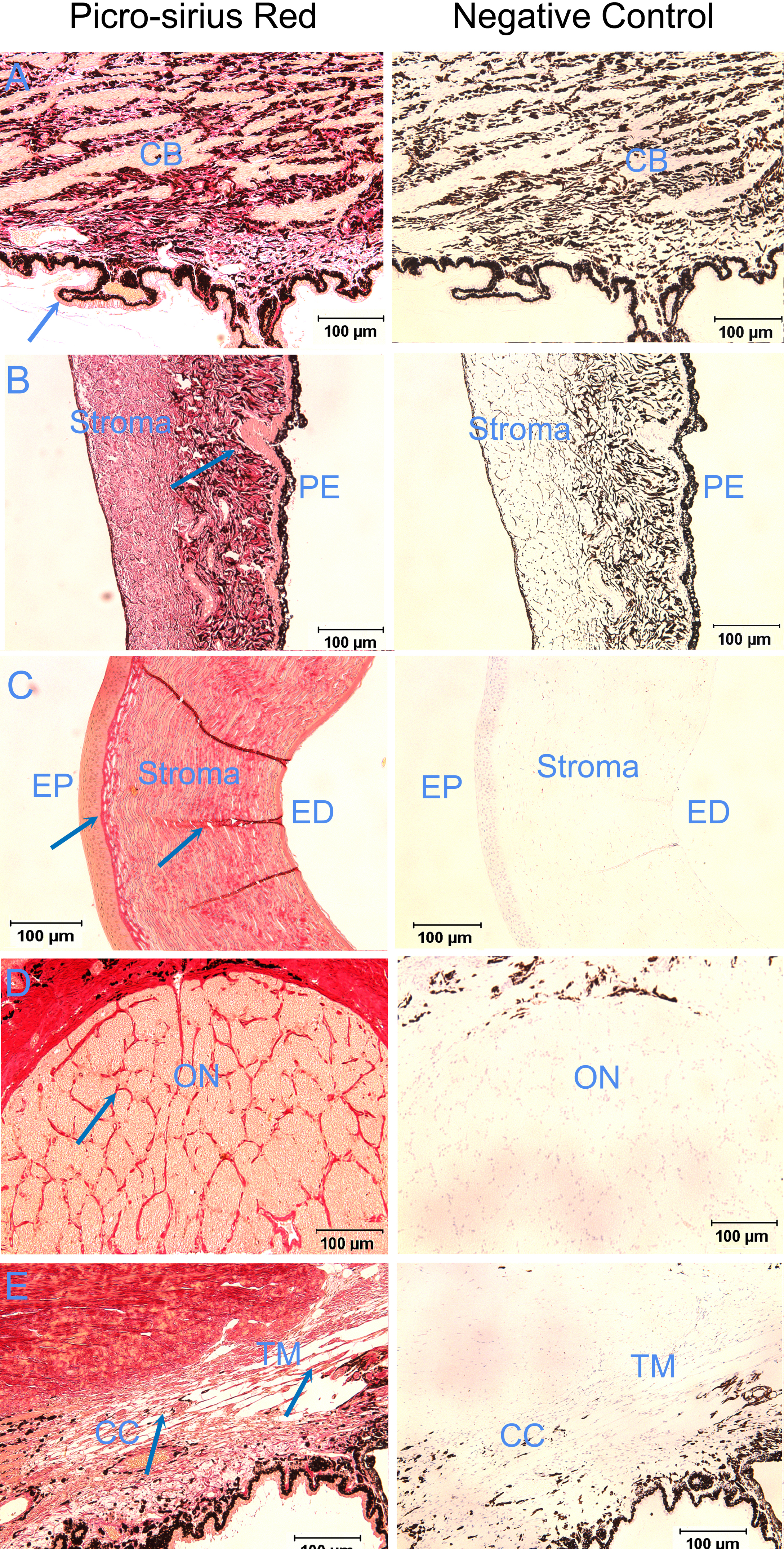
 Figure 5 of
Ahram, Mol Vis 2014; 20:497-510.
Figure 5 of
Ahram, Mol Vis 2014; 20:497-510.